 Back
BackMod 5 pdf
Study Guide - Smart Notes
 Tailored notes based on your materials, expanded with key definitions, examples, and context.
Peripheral Nervous System (PNS) Overview
Divisions and Functional Organization
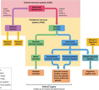
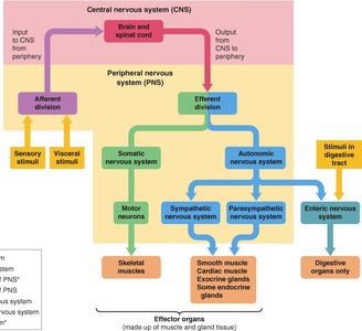
The peripheral nervous system (PNS) connects the central nervous system (CNS) to limbs and organs, facilitating communication between the body and the brain. It is divided into afferent (sensory) and efferent (motor) divisions, each with specialized roles.
Afferent Division: Transmits sensory and visceral stimuli to the CNS.
Efferent Division: Carries signals from the CNS to effector organs via the somatic and autonomic nervous systems.
Somatic Nervous System: Controls voluntary movements via motor neurons targeting skeletal muscles.
Autonomic Nervous System: Regulates involuntary functions, subdivided into sympathetic, parasympathetic, and enteric systems.
Neuronal Pathways
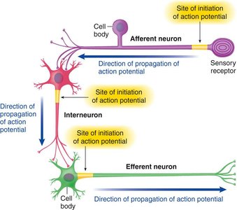
Neurons transmit information through action potentials. The pathway typically involves sensory receptors, afferent neurons, interneurons, and efferent neurons.
Afferent Neurons: Carry sensory information from receptors to the CNS.
Interneurons: Integrate information within the CNS.
Efferent Neurons: Transmit signals from the CNS to effectors (muscles/glands).
Mechanisms for Sensing the Environment
Types of Sensory Receptors
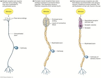
Specialized receptors detect environmental changes and convert them into neural signals. These include:
Photoreceptors: Detect light (vision).
Mechanoreceptors: Sense mechanical energy (touch, pressure, vibration).
Thermoreceptors: Respond to temperature changes.
Osmoreceptors: Monitor solute concentrations.
Nociceptors: Detect tissue damage (pain).
Receptor Activation and Signal Transduction
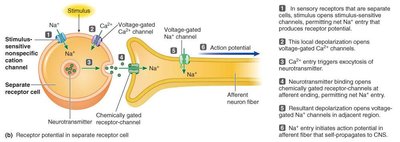
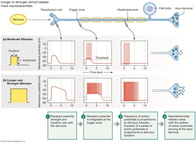
Activation of sensory receptors leads to changes in membrane permeability, often resulting in an influx of Na+ ions and the generation of a receptor potential. If the receptor is a separate cell, neurotransmitter release triggers an action potential in the afferent neuron.
Graded Potentials and Action Potentials
The magnitude of the graded (receptor) potential determines the frequency of action potentials generated. Stronger or longer stimuli produce higher frequencies of action potentials, leading to increased neurotransmitter release at synapses.
Receptor Adaptation
Tonic vs. Phasic Receptors
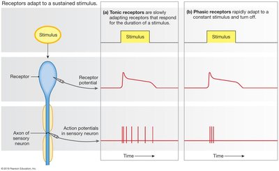
Receptors adapt to sustained stimuli in different ways:
Tonic Receptors: Adapt slowly and provide continuous information about a stimulus (e.g., muscle stretch receptors, joint proprioceptors).
Phasic Receptors: Adapt rapidly and signal changes in stimulus intensity (e.g., touch receptors).
Types of Touch Receptors
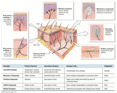
Touch receptors in the skin include free nerve endings, Meissner's corpuscles, Pacinian corpuscles, Ruffini corpuscles, and Merkel receptors. Each type is specialized for different aspects of touch, pressure, and vibration.
Receptive Fields and Sensory Acuity
Receptive Field Size and Sensitivity
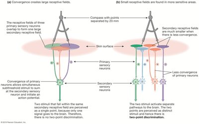
The receptive field is the area monitored by a single sensory neuron. Smaller receptive fields allow for greater acuity and discrimination of stimuli, while larger fields reduce sensitivity.
Sensory Homunculus
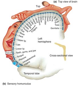
The sensory homunculus is a representation of the body mapped onto the somatosensory cortex, illustrating the relative sensitivity of different regions. Areas with more sensory receptors (e.g., lips, fingertips) occupy larger cortical regions.
Lateral Inhibition
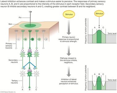
Lateral inhibition enhances contrast and sharpens sensory perception by inhibiting neighboring neurons, making it easier to localize stimuli.
Pain and Nociception
Nociceptors and Pain Types
Nociceptors are specialized receptors for pain that do not adapt to sustained stimuli. They are classified as:
Mechanical Nociceptors: Respond to cutting, crushing, or pinching.
Thermal Nociceptors: Detect extreme temperatures.
Polymodal Nociceptors: Respond to damaging stimuli, including chemicals released during inflammation.
Characteristics of Pain
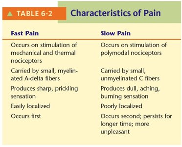
Fast Pain | Slow Pain |
|---|---|
Occurs on stimulation of mechanical and thermal nociceptors | Occurs on stimulation of polymodal nociceptors |
Carried by small, myelinated A-delta fibers | Carried by small, unmyelinated C fibers |
Produces sharp, pricking sensation | Produces dull, aching, burning sensation |
Easily localized | Poorly localized |
Occurs first | Occurs second; persists longer; more unpleasant |
Chemical Mediators and Pain Pathways
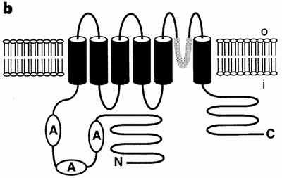
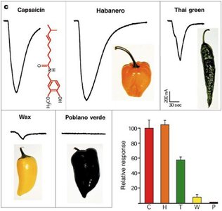
Substances such as bradykinin, substance P, and glutamate enhance pain perception and activate ascending pain pathways. The capsaicin receptor (TRPV1) is a non-selective cation channel activated by capsaicin and noxious heat.
Pain Modulation and Opioids
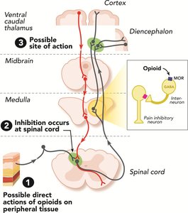
Pain signals can be modulated at multiple levels, including the spinal cord and brain. Opioids inhibit pain transmission by acting on specific receptors, reducing the perception of pain.
Special Senses
Vision: Eye Structure and Protection
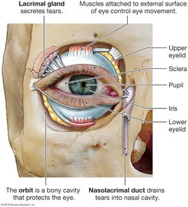
The eye is protected by the bony orbit, eyelids, eyelashes, and tears produced by the lacrimal glands. Tears lubricate, cleanse, and protect the eye from infection.
Eye Anatomy and Chambers
The eye consists of several layers and chambers:
Sclera: Tough outer layer (white of the eye).
Cornea: Transparent anterior layer for light entry.
Choroid: Vascular layer nourishing the retina.
Retina: Contains photoreceptors and neural layers.
Anterior and Posterior Chambers: Filled with aqueous and vitreous humor, respectively.
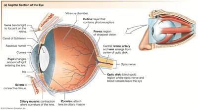
Focusing and Accommodation
The lens is convex to focus light on the retina. Accommodation involves the ciliary muscles adjusting lens shape for near or distant vision.
Photoreceptor Function
Photoreceptors (rods and cones) absorb light and initiate visual signals. In darkness, photoreceptors are depolarized and release glutamate; in light, they hyperpolarize and reduce glutamate release.
Special Senses: Auditory, Olfactory, and Gustatory Systems
Auditory System
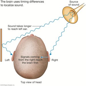
Sound waves are detected by mechanoreceptors (hair cells) in the cochlea. The auditory system identifies and localizes sounds using timing and intensity differences between ears.
Olfactory System
Olfactory receptors are specialized neurons in the nasal mucosa. They detect odorants and transmit signals to the brain for odor perception.
Gustatory System
Taste receptors detect five primary tastes: salty, sour, sweet, bitter, and umami. Each taste is triggered by specific chemicals or ions interacting with receptor cells on the tongue.
Example: Salty taste is primarily due to NaCl, while umami is evoked by glutamate.
Additional info: The mechanisms of taste transduction differ for each taste modality, involving ion channels or G-protein-coupled receptors.
