 Back
BackPeripheral Nervous System: Functional and Structural Organization
Study Guide - Smart Notes
 Tailored notes based on your materials, expanded with key definitions, examples, and context.
Peripheral Nervous System (PNS): Functional and Structural Organization
Overview of the PNS
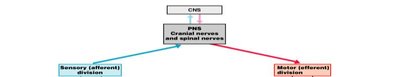
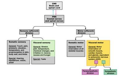
The Peripheral Nervous System (PNS) connects the Central Nervous System (CNS) to the rest of the body, facilitating communication between the brain, spinal cord, and peripheral tissues. The PNS is divided into sensory (afferent) and motor (efferent) divisions, each with specialized functions.
Sensory (afferent) division: Transmits sensory information from receptors to the CNS.
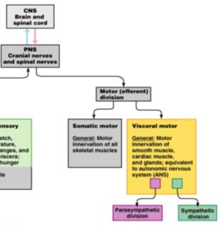
Motor (efferent) division: Sends motor commands from the CNS to effectors (muscles and glands).
Functional Subdivisions of the PNS
The PNS is further subdivided based on the type of information processed and the target tissues.
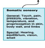
Somatic Sensory: General senses (touch, pain, pressure, vibration, temperature, proprioception) and special senses (hearing, equilibrium, vision, smell).
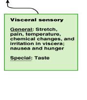
Visceral Sensory: General senses (stretch, pain, temperature, chemical changes, irritation in viscera, nausea, hunger) and special sense (taste).
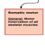
Somatic Motor: Motor innervation of all skeletal muscles.
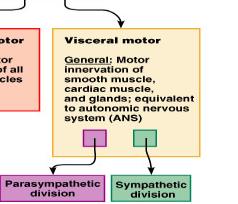
Visceral Motor: Motor innervation of smooth muscle, cardiac muscle, and glands; equivalent to the autonomic nervous system (ANS), which includes the parasympathetic and sympathetic divisions.
Structural Organization of the PNS
Components of the PNS
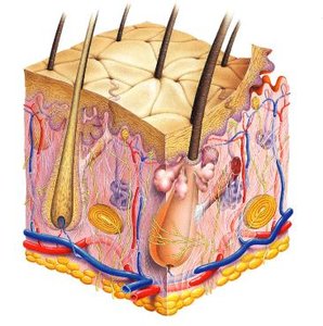
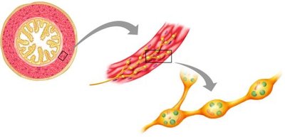
The structural organization of the PNS includes nerves, ganglia, sensory receptors, and motor endings. These components work together to transmit signals between the CNS and peripheral tissues.
Nerves: Bundles of axons that carry information to and from the CNS.
Dorsal root ganglia: Clusters of sensory neuron cell bodies located near the spinal cord.
Sensory receptors: Specialized structures that detect stimuli.
Motor endings: Structures where motor neurons innervate target tissues.
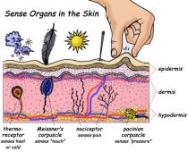
Classification of Sensory Receptors
Sensory receptors are classified by structure, location, and function. They detect various types of stimuli and initiate action potentials in sensory neurons.
Mechanoreceptors: Respond to mechanical forces (pressure, vibration, touch).
Thermoreceptors: Detect temperature changes.
Chemoreceptors: Respond to chemical stimuli (e.g., taste, blood chemistry).
Photoreceptors: Detect light (primarily in the retina).
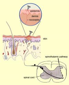
Nociceptors: Respond to pain (mechanical, thermal, or chemical stimuli).
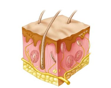
Mechanoreceptors and Proprioceptors
Mechanoreceptors in the Skin
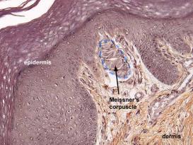
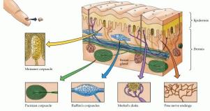
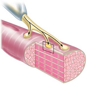
Mechanoreceptors are specialized to detect physical distortion, such as pressure and vibration. Examples include Meissner's corpuscles (light touch) and root hair plexuses (hair movement).
Meissner's corpuscles: Located just beneath the epithelium in hairless skin, lips, and fingertips; sensitive to light touch.
Root hair plexus: Free nerve endings wrapped around hair follicles, responding to hair movement.
Proprioceptors
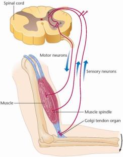
Proprioceptors are mechanoreceptors located in muscles, tendons, and joint capsules. They detect stretch and provide information about body position and movement, which is essential for coordination.
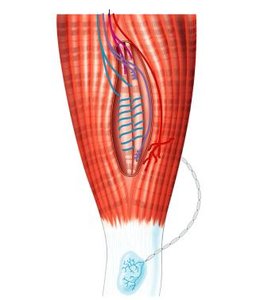
Muscle spindle: Detects stretch in skeletal muscle fibers.
Golgi tendon organ: Responds to tension in tendons.
Other Sensory Receptors
Thermoreceptors
Thermoreceptors are free nerve endings distributed throughout the body that respond to temperature changes.
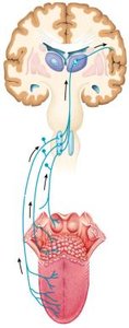
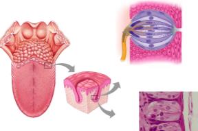
Chemoreceptors
Chemoreceptors respond to specific chemicals, such as those involved in taste and blood chemistry. Taste buds are an example of chemoreceptors.
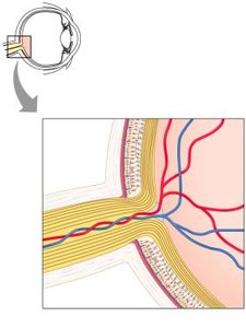
Photoreceptors
Photoreceptors are specialized cells in the retina that respond to light, enabling vision.
Nociceptors
Nociceptors are free nerve endings that respond to potentially damaging stimuli, such as excessive pressure, extreme temperatures, or chemicals released from damaged cells. They are responsible for the sensation of pain.
Motor Response and Neuromuscular Junction
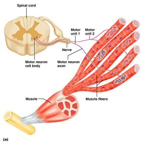
Motor Units
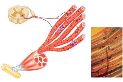
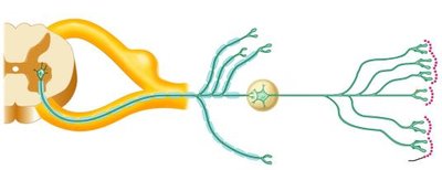
A motor unit consists of a single somatic motor neuron and all the muscle fibers it innervates. This organization allows for precise control of muscle contraction.
Neuromuscular Junction
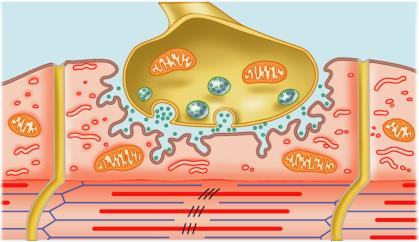
The neuromuscular junction is the site where a motor neuron communicates with a skeletal muscle fiber. Neurotransmitters, such as acetylcholine, are released to initiate muscle contraction.
Innervation of Smooth Muscle
Motor neurons also innervate smooth muscle cells, releasing neurotransmitters such as acetylcholine or norepinephrine to regulate contraction.
Cranial and Spinal Nerves
Cranial Nerves
There are 12 pairs of cranial nerves that emerge from the brain and innervate structures in the head and neck.

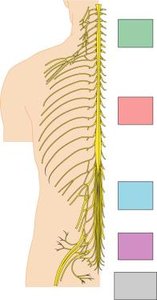
Spinal Nerves
There are 31 pairs of spinal nerves, categorized as cervical, thoracic, lumbar, sacral, and coccygeal. These nerves innervate the body and are essential for sensory and motor function.
Cervical: 8 pairs
Thoracic: 12 pairs
Lumbar: 5 pairs
Sacral: 5 pairs
Coccygeal: 1 pair
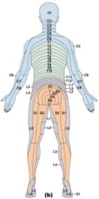
Dermatomes
Dermatomes are regions of skin innervated by specific spinal nerves. Mapping dermatomes is clinically useful for diagnosing spinal cord injuries.
Nerve Plexuses
Nerve plexuses are networks of nerves that supply specific regions of the body. The main plexuses are cervical, brachial, lumbar, and sacral.

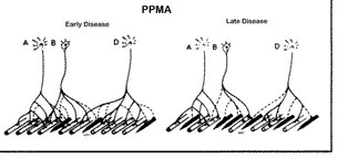
Polio and Motor Neuron Disease
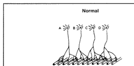
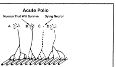
Polio Virus
Polio is a viral disease that targets motor neurons, leading to paralysis. The extent of paralysis depends on which motor neurons are affected.
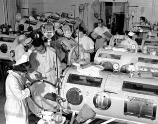

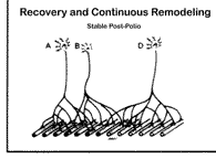
Motor Neuron Recovery and Remodeling
Surviving motor neurons may develop axonal sprouts to reinnervate muscle fibers that lost their original innervation. Over time, these connections may deteriorate due to overuse.
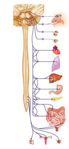
Autonomic Nervous System (ANS)
Overview of the ANS
The Autonomic Nervous System (ANS) is a division of the PNS responsible for involuntary motor responses. It innervates smooth muscle, cardiac muscle, and glands, regulating functions such as digestion, blood pressure, urination, heart rate, and glandular secretion.
Parasympathetic and Sympathetic Divisions
The ANS consists of two divisions that balance each other:
Parasympathetic Division: Oversees resting and digesting functions, conserves energy, and has a localized effect.
Sympathetic Division: Mobilizes the body in extreme situations (fight, fright, or flight), requires more energy, and has a widespread effect.
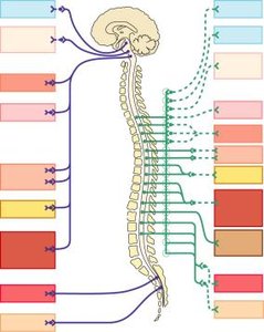
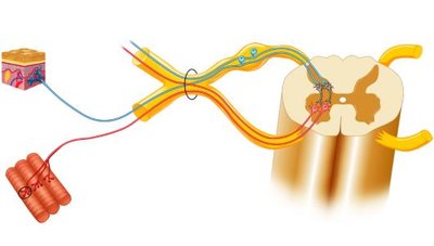
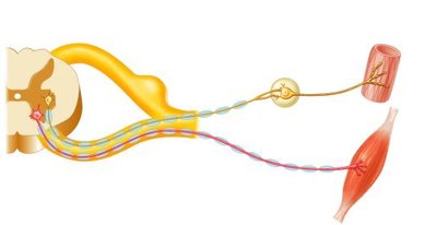
Structural Differences: Somatic vs. Autonomic Motor Pathways
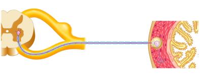
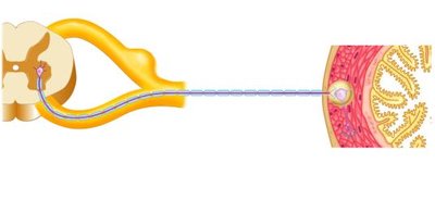
Somatic motor neurons have a single neuron running from the spinal cord to skeletal muscle. Visceral motor neurons (ANS) use two neurons: preganglionic and postganglionic, which synapse in an autonomic ganglion.
Neurotransmitters in the ANS
Different neurotransmitters are released depending on the division and location:
Somatic motor neuron: Acetylcholine
Parasympathetic pathway: Acetylcholine at both synapses
Sympathetic pathway: Acetylcholine at the ganglion, norepinephrine at the target tissue
Parasympathetic Pathway
Parasympathetic preganglionic fibers are long with limited branching, and postganglionic fibers are short. The autonomic ganglion is near or within the target tissue.

Sympathetic Pathway
Sympathetic preganglionic fibers are short with extensive branching, and postganglionic fibers are long. The autonomic ganglion is near the spinal cord.

Summary Table: Functional Divisions of the PNS
Division | General Function | Special Function |
|---|---|---|
Somatic Sensory | Touch, pain, pressure, vibration, temperature, proprioception in skin, body wall, limbs | Hearing, equilibrium, vision, smell |
Visceral Sensory | Stretch, pain, temperature, chemical changes, irritation in viscera; nausea, hunger | Taste |
Somatic Motor | Motor innervation of all skeletal muscles | None |
Visceral Motor (ANS) | Motor innervation of smooth muscle, cardiac muscle, glands | Parasympathetic and Sympathetic divisions |
