 Back
BackPeripheral Nervous System Physiology: Structure, Function, and Reflexes
Study Guide - Smart Notes
 Tailored notes based on your materials, expanded with key definitions, examples, and context.
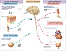
Peripheral Nervous System (PNS) Overview
Divisions of the PNS
The Peripheral Nervous System (PNS) connects the Central Nervous System (CNS) to limbs and organs, serving as a communication relay. It is divided into sensory (afferent) and motor (efferent) divisions, each with distinct roles in transmitting information.
Sensory (Afferent) Division: Carries sensory information from receptors to the CNS.
Motor (Efferent) Division: Transmits motor commands from the CNS to effectors (muscles and glands).
Somatic Nervous System: Controls voluntary movements via skeletal muscles.
Autonomic Nervous System (ANS): Regulates involuntary functions, targeting smooth muscle, cardiac muscle, and glands.
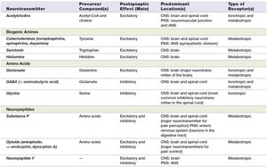
Major Neurotransmitters in the PNS
Types and Functions
Neurotransmitters are chemical messengers that transmit signals across synapses. They can be classified by their chemical structure and function, influencing excitatory or inhibitory responses in target cells.
Acetylcholine (ACh): Main neurotransmitter at neuromuscular junctions and autonomic ganglia; usually excitatory.
Biogenic Amines: Includes norepinephrine, epinephrine, dopamine, serotonin, and histamine; involved in mood, arousal, and autonomic regulation.
Amino Acids: Glutamate (excitatory), GABA and glycine (inhibitory); major neurotransmitters in the CNS.
Neuropeptides: Substance P, opioids, neuropeptide Y; modulate pain, stress, and other functions.
Neurotransmitter | Precursor | Main Effect | Location | Receptor Type |
|---|---|---|---|---|
Acetylcholine | Acetyl-CoA + choline | Excitatory | CNS, PNS | Ionotropic/Metabotropic |
Norepinephrine | Tyrosine | Excitatory/Inhibitory | CNS, PNS | Metabotropic |
Serotonin | Tryptophan | Excitatory/Inhibitory | CNS | Metabotropic |
GABA | Glutamate | Inhibitory | CNS | Ionotropic/Metabotropic |
Substance P | Amino acids | Excitatory | CNS, PNS | Metabotropic |
Neuropeptide Y | - | Excitatory/Inhibitory | CNS, PNS | Metabotropic |
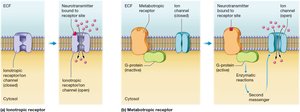
Types of Neurotransmitter Receptors
Ionotropic vs. Metabotropic Receptors
Neurotransmitter receptors determine the cellular response to neurotransmitters. They are classified as:
Ionotropic Receptors: Ligand-gated ion channels that mediate fast synaptic transmission by directly altering membrane potential.
Metabotropic Receptors: G-protein-coupled receptors that initiate slower, longer-lasting effects via second messenger systems.
Example: Nicotinic acetylcholine receptors are ionotropic, while muscarinic acetylcholine receptors are metabotropic.
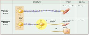
Somatic vs. Autonomic Motor Divisions
Structural and Functional Differences
The motor division of the PNS is divided into somatic and autonomic branches, each with unique pathways and targets.
Somatic Motor Division:
One neuron from CNS to skeletal muscle
No ganglia
Releases acetylcholine (ACh) at the target
Primarily voluntary control
Autonomic Motor Division:
Two-neuron pathway (preganglionic and postganglionic)
Ganglia present
Releases ACh or norepinephrine (NE) at the target
Targets smooth muscle, cardiac muscle, and glands
Involuntary control
Preganglionic vs. Postganglionic Autonomic Neurons
Pathways in the Autonomic Nervous System
Autonomic pathways consist of two neurons:
Preganglionic Neuron: Cell body in the CNS; axon projects to an autonomic ganglion.
Postganglionic Neuron: Cell body in the ganglion; axon projects to the target organ.
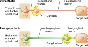
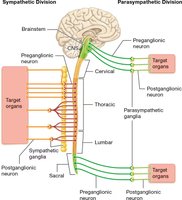
Sympathetic and Parasympathetic Motor Neurons
Organization and Function
The autonomic nervous system is divided into sympathetic and parasympathetic branches, each with distinct anatomical origins and effects.
Sympathetic Division: Originates from thoracic and lumbar spinal cord; prepares body for 'fight or flight' responses.
Parasympathetic Division: Originates from brainstem and sacral spinal cord; promotes 'rest and digest' activities.
Adrenergic and Cholinergic Receptor Subtypes
Sympathetic and Parasympathetic Effects
Autonomic neurotransmitters act on specific receptor subtypes to produce varied physiological effects.
Adrenergic Receptors (Sympathetic):
⍺1: Vasoconstriction, increases blood pressure
⍺2: Inhibits neurotransmitter release (feedback control)
β1: Increases heart rate and contractility
β2: Bronchodilation and vasodilation (skeletal muscle)
β3: Lipolysis (fat breakdown)
Cholinergic Receptors (Parasympathetic):
Nicotinic (N): Fast synaptic transmission; found in autonomic ganglia and neuromuscular junctions
Muscarinic (M): Slower, longer-lasting effects; found on parasympathetic target organs
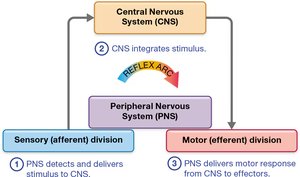
Reflex Components and Homeostasis
Structure of a Reflex Arc
Reflexes are rapid, automatic responses to stimuli that help maintain homeostasis. A typical reflex arc includes:
Receptor: Detects a stimulus
Sensory (Afferent) Neuron: Carries information to the CNS
Integration Center: Processes information (spinal cord or brain)
Motor (Efferent) Neuron: Carries response signal
Effector: Muscle or gland that produces the response
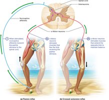
Monosynaptic and Polysynaptic Reflexes
Types of Reflex Pathways
Monosynaptic Reflex:
One synapse between sensory and motor neuron
Fastest reflex pathway
Example: Stretch (knee-jerk) reflex
Function: Maintains muscle length and posture
Polysynaptic Reflex:
Two or more synapses with interneurons
Slower but more complex responses
Example: Withdrawal reflex
Function: Coordinates multiple muscles for protection
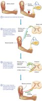
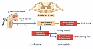
Patellar Tendon (Knee-Jerk) Reflex Pathway
Example of a Monosynaptic Reflex
The patellar tendon reflex is a classic example of a monosynaptic reflex, used clinically to assess nervous system function.
Tap on patellar tendon stretches quadriceps muscle
Muscle spindle (receptor) activates sensory neuron
Sensory neuron synapses directly with motor neuron in spinal cord
Motor neuron stimulates quadriceps to contract, causing leg extension
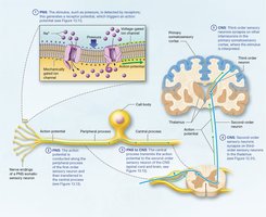
Major Sensory Receptors and Their Stimuli
Types of Sensory Receptors
Sensory receptors detect specific types of stimuli and convert them into neural signals.
Mechanoreceptors: Touch, pressure, vibration, stretch
Thermoreceptors: Temperature changes
Chemoreceptors: Chemicals (taste, smell, blood chemistry)
Baroreceptors: Pressure changes (e.g., blood pressure)
Osmoreceptors: Osmotic pressure (fluid balance)
Proprioceptors: Body position and movement
Nociceptors: Pain or tissue damage
Receptive Field & Two-Point Discrimination
Spatial Resolution of Sensory Neurons
The receptive field is the area where a sensory neuron can detect a stimulus. The size of receptive fields affects tactile resolution and the ability to distinguish two points.
Small Receptive Fields: High tactile precision (e.g., fingertips)
Large Receptive Fields: Lower tactile precision (e.g., back, thighs)
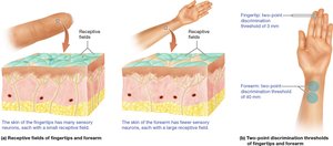
First-Order & Second-Order Neurons and Two-Point Discrimination
Neural Pathways for Sensory Processing
First-order neurons detect stimuli at the skin and send signals to the spinal cord or brainstem. Second-order neurons carry the signal to the thalamus. The precision of two-point discrimination depends on the size and mapping of receptive fields.
Fingertips: Many first-order neurons with small receptive fields → high tactile precision
Back: Fewer first-order neurons with large receptive fields → lower tactile precision
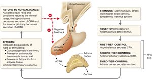
Sympathetic Nervous System & Adrenal Medulla
Role in Stress Response
The adrenal medulla acts as a modified sympathetic ganglion. Sympathetic neurons stimulate the adrenal medulla to release epinephrine and norepinephrine into the bloodstream, producing a widespread, longer-lasting sympathetic response.
Stress Responses: SAM and HPA Pathways
Acute and Chronic Stress Mechanisms
Sympathetic-Adrenal-Medullary (SAM) Response:
Fast response (seconds)
Sympathetic activation → adrenal medulla releases epinephrine
Effects: Increased heart rate, blood pressure, bronchodilation, energy mobilization
Prepares body for immediate action
Hypothalamic-Pituitary-Adrenal (HPA) Response:
Slower response (minutes to hours)
Hypothalamus releases CRH → Pituitary releases ACTH → Adrenal cortex releases cortisol
Effects: Maintains blood glucose, suppresses immune response, supports long-term stress adaptation
Eustress vs. Distress
Eustress: Positive, beneficial stress that improves focus and performance
Distress: Negative, harmful stress that impairs function and health
