 Back
BackPeripheral Nervous System (PNS): Structure, Function, and Organization
Study Guide - Smart Notes
 Tailored notes based on your materials, expanded with key definitions, examples, and context.
Peripheral Nervous System (PNS)
Overview of the Peripheral Nervous System
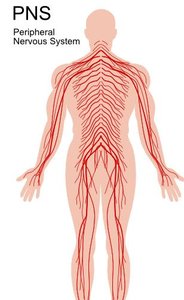
The Peripheral Nervous System (PNS) consists of all neural structures outside the brain and spinal cord. It serves as a communication network linking the Central Nervous System (CNS) to the limbs and organs, thus connecting the CNS to the external environment and the rest of the body.
Cranial nerves: 12 pairs, branch off the brainstem.
Spinal nerves: 31 pairs, branch off the spinal cord.
Includes sensory receptors, afferent and efferent nerves, associated ganglia, and motor endings.
Functional Organization of the PNS
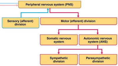
The PNS is divided into sensory (afferent) and motor (efferent) divisions. The motor division is further subdivided into the somatic (voluntary) and autonomic (involuntary) nervous systems. The autonomic system is split into sympathetic and parasympathetic divisions.
Sensory (afferent) division: Carries information toward the CNS.
Motor (efferent) division: Carries information away from the CNS to muscles and glands.
Somatic nervous system: Voluntary control of skeletal muscles.
Autonomic nervous system (ANS): Involuntary control of smooth muscle, cardiac muscle, and glands.
Sympathetic division: Prepares the body for stress ('fight or flight').
Parasympathetic division: Promotes rest and digestion ('rest and digest').
Sensory Receptors and Sensation
Types and Classification of Sensory Receptors
Sensory receptors are specialized to respond to changes in the environment (stimuli). Activation of these receptors triggers graded potentials that may lead to nerve impulses sent to the CNS, where sensation and perception occur.
Mechanoreceptors: Respond to mechanical force (touch, pressure, vibration, stretch).
Thermoreceptors: Detect temperature changes.
Photoreceptors: Respond to light (e.g., retina of the eye).
Chemoreceptors: Detect chemicals (taste, smell, changes in blood chemistry).
Nociceptors: Respond to potentially damaging stimuli (pain).
Pain Perception and Nociceptors
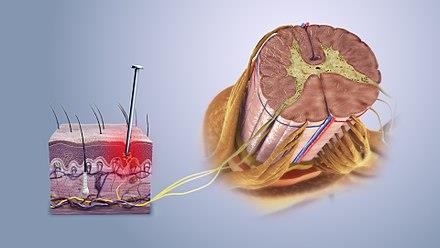
Nociceptors are sensory receptors that detect pain, signaling potential tissue damage. Pain signals travel from the site of injury through the spinal cord to the brain, where they are processed and perceived. Pain perception (threshold) is similar among individuals, but pain tolerance can vary and may be influenced by psychological factors or training.
Visceral pain: Originates from internal organs.
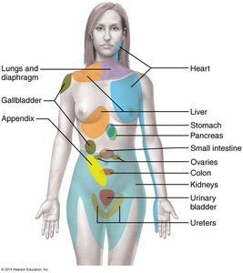
Referred pain: Pain perceived at a location other than the site of the painful stimulus (e.g., left arm pain during a heart attack).
Classification by Location
Exteroceptors: Sensitive to external stimuli (skin, vision, hearing, smell, taste, equilibrium).
Interoceptors (Visceroceptors): Respond to internal stimuli (pain, hunger, thirst, stretch).
Proprioceptors: Located in muscles, tendons, joints; inform the brain about body position and movement.
Nerves and Associated Ganglia
Structure and Classification of Nerves
A nerve is a cordlike organ of the PNS, consisting of bundles of myelinated and unmyelinated axons enclosed by connective tissue. Nerves are classified as cranial or spinal based on their origin and as sensory, motor, or mixed based on the direction of impulse transmission.
Mixed nerves: Contain both sensory and motor fibers; impulses travel both to and from the CNS.
Sensory (afferent) nerves: Carry impulses only toward the CNS.
Motor (efferent) nerves: Carry impulses only away from the CNS.
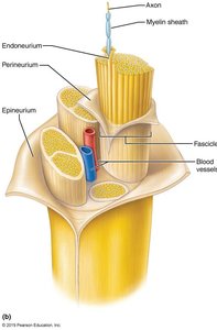
Ganglia
Ganglia are clusters of neuron cell bodies in the PNS. They are associated with either sensory (afferent) or autonomic (efferent) nerve fibers.
Dorsal root ganglia: Contain cell bodies of sensory neurons (somatic).
Autonomic ganglia: Contain cell bodies of autonomic motor neurons (visceral).
Regeneration of Nerve Fibers
Regeneration in the PNS
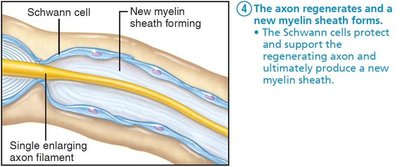
Unlike the CNS, peripheral axons can regenerate if the cell body is intact and the damage is not severe. The process involves several steps:
Axon fragments and myelin sheaths distal to injury degenerate (Wallerian degeneration).
Macrophages clean up debris; Schwann cells proliferate.
Axon filaments grow through a regeneration tube formed by Schwann cells.
The axon regenerates, and a new myelin sheath forms.
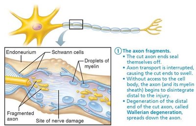
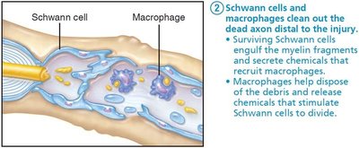
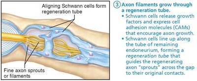
Spinal Nerves and Their Organization
Spinal Nerve Structure and Distribution
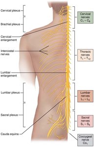
There are 31 pairs of spinal nerves, all of which are mixed nerves. They are named according to their point of origin from the spinal cord and supply all body parts except the head and part of the neck.
8 pairs of cervical nerves (C1–C8)
12 pairs of thoracic nerves (T1–T12)
5 pairs of lumbar nerves (L1–L5)
5 pairs of sacral nerves (S1–S5)
1 pair of coccygeal nerves (C0)
Spinal Cord Cross Section
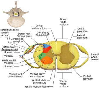
The spinal cord consists of white matter (myelinated axons) on the outside and gray matter (dendrites and cell bodies) on the inside. The dorsal horn receives sensory (afferent) information, while the ventral horn sends motor (efferent) output.
Spinal Nerve Roots and Branches
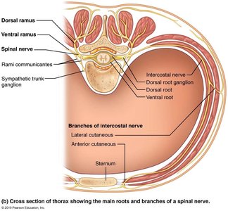
Each spinal nerve connects to the spinal cord via two roots:
Ventral roots: Motor (efferent) fibers from ventral horn motor neurons.
Dorsal roots: Sensory (afferent) fibers from sensory neurons in dorsal root ganglia.
After exiting the vertebral foramen, spinal nerves divide into three branches:
Dorsal ramus: Supplies posterior body trunk.
Ventral ramus: Supplies the rest of the trunk and limbs.
Meningeal branch: Reenters vertebral canal to innervate meninges and blood vessels.
Nerve Plexuses
Organization and Function of Nerve Plexuses
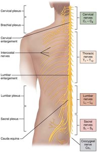
All ventral rami except T2–T12 form interlacing nerve networks called nerve plexuses. These are found in the cervical, brachial, lumbar, and sacral regions. Plexuses allow fibers from different spinal nerves to combine and redistribute, ensuring that each limb muscle is innervated by more than one spinal nerve, which provides redundancy and protection from paralysis if one nerve is damaged.
Somatic and Autonomic Nervous Systems
Somatic Nervous System
The somatic nervous system is responsible for voluntary control of body movements via skeletal muscles. It includes both sensory (afferent) and motor (efferent) nerves and is involved in conscious activities and reflex arcs.
Autonomic Nervous System: Sympathetic vs. Parasympathetic
The autonomic nervous system (ANS) regulates involuntary physiological processes, including heart rate, blood pressure, respiration, digestion, and sexual arousal. It has two main divisions with opposing functions:
Sympathetic division: Prepares the body for stress ('fight or flight').
Parasympathetic division: Promotes restorative processes ('rest and digest').
