 Back
BackPeripheral Nervous System (PNS): Structure, Function, and Reflexes
Study Guide - Smart Notes
 Tailored notes based on your materials, expanded with key definitions, examples, and context.
Peripheral Nervous System (PNS)
Overview of the PNS
The Peripheral Nervous System (PNS) connects the central nervous system (CNS) to limbs and organs, serving as a communication relay between the brain, spinal cord, and the rest of the body. It is essential for sensation, movement, and reflexes.
Key Definitions
Sensation: The awareness of changes in the internal and external environment, detected by sensory receptors.
Perception: The conscious interpretation of sensory stimuli, allowing us to understand and respond to our environment.
Nerves: Bundles of axons in the PNS that transmit sensory and motor information.
Ganglia: Collections of neuron cell bodies located outside the CNS, often serving as relay points in neural pathways.
Types of Sensory Receptors
By Stimulus Type
Mechanoreceptors: Respond to mechanical force (touch, pressure, vibration).
Thermoreceptors: Detect changes in temperature.
Photoreceptors: Respond to light (e.g., in the retina).
Chemoreceptors: Detect chemicals in solution (taste, smell, blood chemistry).
Nociceptors: Respond to potentially damaging stimuli that result in pain.
By Structural Complexity
Simple receptors: General senses (touch, pain, temperature).
Complex receptors: Special senses (vision, hearing, equilibrium, taste, smell).
Adaptation of Sensory Receptors
Adaptation: A decrease in receptor sensitivity with constant stimulus.
Phasic receptors: Fast-adapting; signal the beginning or end of a stimulus (e.g., pressure, touch).
Tonic receptors: Slow-adapting; provide a sustained response (e.g., pain, proprioceptors).
Pain
Somatic pain: Originates from skin, muscles, and joints; usually well localized.
Visceral pain: Originates from internal organs; often poorly localized and can be referred to other areas.
Referred pain: Pain perceived at a location other than the site of the painful stimulus, due to shared neural pathways.
Structure of a PNS Nerve
Endoneurium: Surrounds individual axons.
Perineurium: Surrounds bundles of axons (fascicles).
Epineurium: Encloses the entire nerve.
Nerve Regeneration After Damage
Peripheral nerves can regenerate if the cell body remains intact. Schwann cells play a key role in guiding axonal regrowth, but regeneration is slow and often incomplete.
Cranial Nerves
Names and Functions
There are 12 pairs of cranial nerves, each with specific sensory, motor, or mixed functions. Some also carry parasympathetic (PS*) fibers.
Cranial Nerve | Sensory Function | Motor Function | PS* Fibers |
|---|---|---|---|
I Olfactory | Yes (smell) | No | No |
II Optic | Yes (vision) | No | No |
III Oculomotor | No | Yes | Yes |
IV Trochlear | No | Yes | No |
V Trigeminal | Yes (general sensation) | Yes | No |
VI Abducens | No | Yes | No |
VII Facial | Yes (taste) | Yes | Yes |
VIII Vestibulocochlear | Yes (hearing and balance) | No | No |
IX Glossopharyngeal | Yes (taste) | Yes | Yes |
X Vagus | Yes (taste) | Yes | Yes |
XI Accessory | No | Yes | No |
XII Hypoglossal | No | Yes | No |
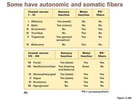
PS* = parasympathetic fibers
Spinal Nerves
There are 31 pairs of spinal nerves, grouped into five regions:
Cervical
Thoracic
Lumbar
Sacral
Coccygeal
Plexuses
Plexus: A network of intersecting nerves. Major plexuses include:
Cervical plexus: Serves the head, neck, and shoulders.
Brachial plexus: Serves the upper limb.
Lumbar plexus: Serves the anterior thigh.
Sacral plexus: Serves the posterior thigh, lower leg, and foot.
Dermatomes
Dermatome: An area of skin supplied by sensory fibers from a single spinal nerve.
Reflexes
Reflex: A rapid, automatic response to a stimulus.
Inborn (intrinsic) reflexes: Present at birth (e.g., knee-jerk reflex).
Learned (acquired) reflexes: Developed through experience (e.g., driving skills).
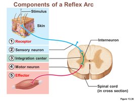
Reflex Arc
The reflex arc is the basic functional unit of the nervous system, consisting of five components:
Receptor: Detects the stimulus.
Sensory neuron: Transmits afferent impulses to the CNS.
Integration center: Processes information (may involve interneurons).
Motor neuron: Conducts efferent impulses to an effector.
Effector: Muscle or gland that responds to the impulse.
Examples of Reflexes
Patellar (knee jerk) reflex: A stretch reflex that helps maintain posture and balance.
Flexor (withdrawal) reflex: Causes withdrawal of a body part from a painful stimulus.
Crossed extensor reflex: Maintains balance when the flexor reflex is initiated in one limb.
Plantar reflex: Stroking the sole of the foot causes downward flexion of the toes.
Babinski’s sign: Abnormal response in adults (toes fan upward), indicating possible CNS damage.
