 Back
BackPeripheral Nervous System, Sensory Receptors, and Special Senses: Study Guide
Study Guide - Smart Notes
 Tailored notes based on your materials, expanded with key definitions, examples, and context.
Peripheral Nervous System (PNS)
Overview of the PNS
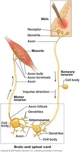
The Peripheral Nervous System (PNS) consists of all neural structures outside the brain and spinal cord. It is responsible for transmitting sensory information to the central nervous system (CNS) and carrying motor commands from the CNS to the body.
Sensory receptors: Specialized cells that detect changes in the environment.
Peripheral nerves and ganglia: Bundles of axons and clusters of neuron cell bodies.
Motor endings: Structures such as axon terminals and motor end plates that connect to effectors (muscles or glands).
Sensory Mechanisms and Neural Integration
From Sensation to Perception
Sensation and perception are critical for survival, allowing organisms to detect and interpret changes in their internal and external environments.
Sensation: Awareness of changes in the environment.
Perception: Conscious integration and interpretation of sensory stimuli by the brain.
Levels of Neural Integration
Sensory systems process information at three basic levels:
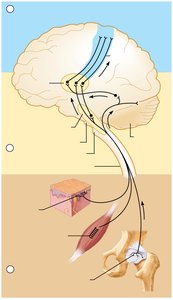
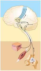
Receptor level: Sensory reception and transduction (conversion of stimulus to graded potentials).
Circuit level: Processing in ascending pathways (transmission via action potentials).
Perceptual level: Processing in cortical sensory areas (conscious perception).
Receptive Fields and Sensory Acuity
Receptive Field Size
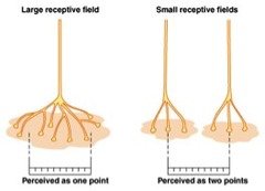
The acuity of sensory perception depends on the size and density of receptive fields. Smaller receptive fields with higher receptor density allow for greater discriminative ability.
Large receptive fields: Lower acuity, perceived as one point.
Small receptive fields: Higher acuity, perceived as two points.
Sensory Adaptation
Receptor Adaptation
Sensory adaptation occurs when a sensory neuron stops sending impulses despite the continued presence of a stimulus. This allows the nervous system to ignore unimportant stimuli and focus on changes.
Phasic receptors: Adapt quickly (e.g., olfaction, light touch).
Tonic receptors: Adapt slowly or not at all (e.g., pain, joint, muscle receptors).
Example: The sensation of clothing fades after a short period due to adaptation.
Classification of Sensory Receptors
By Stimulus Type
Mechanoreceptors: Detect touch, pressure, vibration, stretch, and itch.
Thermoreceptors: Detect changes in temperature.
Photoreceptors: Detect light energy (e.g., retina).
Chemoreceptors: Detect chemicals (e.g., smell, taste, blood chemistry).
Nociceptors: Detect pain-causing stimuli (e.g., extreme heat/cold, pressure, chemicals).
By Location
Somatic receptors: Located throughout the body; detect pressure, temperature, pain, touch, body position, and movement.
Special receptors: Located in specific sense organs; detect taste, smell, hearing, balance, and vision.
Somatic Sensory Receptors and Reflexes
Proprioceptors and Reflexes
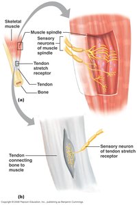
Mechanoreceptors in skeletal muscles, tendons, and joints (proprioceptors) respond to changes in muscle length, tendon tension, and joint position. Somatic reflexes help maintain balance and posture and prevent injury.
Reflex Arc Components
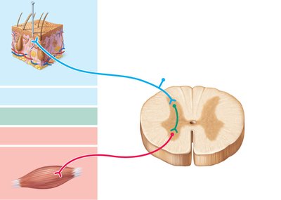
A reflex arc is the neural pathway that mediates a reflex action. It consists of five main components:
Receptor: Detects the stimulus.
Sensory neuron: Transmits the afferent impulse to the CNS.
Integration center: Processes the information (may involve interneurons).
Motor neuron: Conducts efferent impulses to the effector.
Effector: Muscle or gland that responds to the stimulus.
Stretch and Golgi Tendon Reflexes
Stretch Reflexes
Stretch reflexes maintain muscle tone and cause muscle contraction in response to increased muscle length. These reflexes are monosynaptic and ipsilateral.
Example: The patellar (knee-jerk) reflex.
Reciprocal inhibition: Inhibition of antagonist muscles during reflex contraction.
Golgi Tendon Reflexes
Golgi tendon reflexes are polysynaptic and help prevent damage due to excessive muscle tension. They produce muscle relaxation in response to tension and involve reciprocal activation of antagonist muscles.
Special Senses
Overview of Special Senses
Special senses originate from receptors located in specific areas of the body. The five special senses are taste, smell, hearing, balance, and vision.
Taste (Gustation)
Structure and Function of Taste Buds
Taste depends on chemoreceptors found on gustatory epithelial cells within taste buds, primarily located on the papillae of the tongue. Taste buds convert chemical signals from food into action potentials.
Five basic taste modalities: Sweet, sour, salty, bitter, umami (savory).
Most taste receptors: Located on the outer edges of the tongue.
Gustatory Pathway
Tastants bind to chemoreceptors, causing depolarization and neurotransmitter release. Cranial nerves VII, IX, and X carry impulses from taste buds to the medulla, then to the thalamus, and finally to the gustatory cortex in the insula. The limbic system and hypothalamus are also involved in taste perception and emotional responses.
Smell (Olfaction)
Olfactory Receptors and Pathway
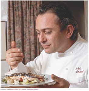
Olfactory chemoreceptors detect more than 1,000 different odorants. Olfactory hairs project into the olfactory epithelium, where odorants dissolve in mucus and bind to receptors, generating impulses.
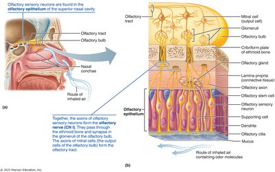
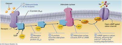
Olfactory receptor cells: Synapse with neurons in the olfactory bulb, where information is partially integrated before being sent to the temporal lobes.
Vision
Light and Photoreceptors
Light is composed of photons that travel in waves. The eyes respond to visible light (400-700 nm), and photoreceptors (rods and cones) detect different wavelengths.
Rods: Dim-light and peripheral vision; more sensitive to light but do not provide clear outlines.
Cones: Bright light and color vision; provide sharp outlines and color discrimination.
Color blindness: Lack of one or more cone cell types.
Visual Pathway
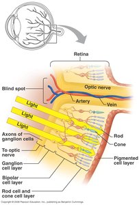
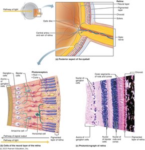
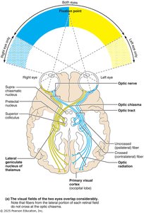
Rods and cones synapse with bipolar cells, which synapse with ganglion cells. Action potentials travel down ganglion cell axons and leave the retina through the optic nerve. At the optic chiasm, fibers cross to the contralateral side and continue via the optic tract to the thalamus and primary visual cortex in the occipital lobe.
Hearing
Sound and Hearing Range
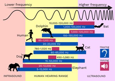
Sounds are waves of compressed air. Sound intensity (energy/amplitude) and loudness (subjective interpretation) are measured in decibels (dB). The human hearing range is 20-20,000 Hz, with other animals capable of hearing higher or lower frequencies.
Anatomy of the Ear
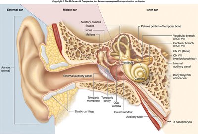
The ear is divided into three main parts:
External ear: Responsible for hearing.
Middle ear: Contains auditory ossicles; responsible for hearing.
Inner ear: Responsible for hearing and balance; contains fluid-filled chambers.
Sound Transduction
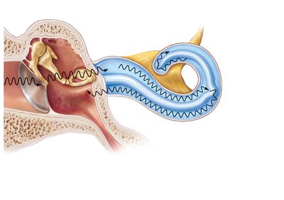
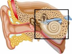
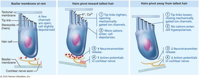
Sound waves travel through the ear, eventually activating vibration-sensitive mechanoreceptors (hair cells) in the cochlea. The vibration produces impulses that travel to the brain for interpretation.
Summary Table: Sensory Receptor Classification
Type | Stimulus Detected | Location | Example |
|---|---|---|---|
Mechanoreceptor | Touch, pressure, vibration, stretch, itch | Skin, muscles, tendons | Muscle spindle, Pacinian corpuscle |
Thermoreceptor | Temperature | Skin | Free nerve endings |
Photoreceptor | Light | Retina | Rods, cones |
Chemoreceptor | Chemicals | Tongue, nose, blood vessels | Taste buds, olfactory epithelium |
Nociceptor | Pain-causing stimuli | Throughout body | Free nerve endings |
Key Equations
Action Potential Frequency:
Sound Intensity (dB):
Points to Remember
Sensation begins with a sensory receptor on a sensory neuron.
Sensory information is sent to the CNS for integration and processing.
Different receptors detect different stimuli and send information to specific CNS areas.
