 Back
BackPeripheral Nervous System, Special Senses, and Muscle Tissue: Study Guide for ANP College Students
Study Guide - Smart Notes
 Tailored notes based on your materials, expanded with key definitions, examples, and context.
Peripheral Nervous System
Cranial Nerves: Location and Function
The cranial nerves are twelve pairs of nerves that emerge directly from the brain and brainstem, each with specific sensory, motor, or mixed functions. They are identified by Roman numerals and names, and their functions range from smell and vision to facial movement and autonomic control.
I Olfactory: Sensory; responsible for smell.
II Optic: Sensory; responsible for vision.
III Oculomotor: Motor; moves the eye and dilates pupils.
IV Trochlear: Motor; innervates extrinsic eye muscles.
V Trigeminal: Both; sensations of head and face, chewing.
VI Abducens: Motor; abducts the eye.
VII Facial: Both; taste, secretion of saliva/tears, facial expression.
VIII Vestibulocochlear: Sensory; hearing and balance.
IX Glossopharyngeal: Both; tongue sensations, saliva secretion, swallowing.
X Vagus: Both; sensations from thoracic/abdominal viscera, parasympathetic control, voice production.
XI Accessory: Motor; shoulder movements, assists in voice production.
XII Hypoglossal: Motor; tongue movements.
Spinal Nerves and Plexuses
Spinal nerves are mixed nerves that originate from the spinal cord, totaling 31 pairs. They are grouped by region and form plexuses that innervate specific body areas.
Cervical: 8 pairs
Thoracic: 12 pairs
Lumbar: 5 pairs
Sacral: 5 pairs
Coccygeal: 1 pair
The anterior and posterior rami of spinal nerves branch to supply motor and sensory innervation to the body.
Cervical Plexus
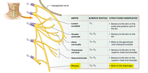
The cervical plexus is formed by the anterior rami of C1–C5 and innervates the neck, shoulder, and diaphragm. The phrenic nerve is especially important for motor control of the diaphragm.
Lesser occipital: Sensory to scalp and ear
Greater auricular: Sensory to skin on ear
Ansa cervicalis: Motor to geniohyoid and infrahyoid muscles
Transverse cervical: Sensory to skin on superior chest/shoulder
Supraclavicular: Sensory to skin on superior chest/shoulder
Phrenic: Motor to the diaphragm
Brachial Plexus
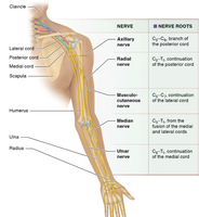
The brachial plexus is formed by the anterior rami of C5–T1 and innervates the upper limb. Key nerves include the median and musculocutaneous nerves, which control forearm and hand muscles.
Axillary: C5–C6; posterior cord
Radial: C5–T1; posterior cord
Musculocutaneous: C5–C7; lateral cord
Median: C6–T1; medial/lateral cord fusion
Ulnar: C8–T1; medial cord
Lumbar Plexus
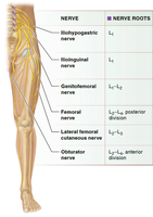
The lumbar plexus is formed by the anterior rami of L1–L4 and innervates the lower abdomen, anterior thigh, and medial leg.
Iliohypogastric: L1
Ilioinguinal: L1
Genitofemoral: L1–L2
Femoral: L2–L4; posterior division
Lateral femoral cutaneous: L2–L3
Obturator: L2–L4; anterior division
Sacral Plexus
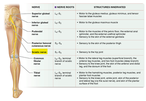
The sacral plexus is formed by the anterior rami of L4–S4 and innervates the pelvis, posterior thigh, and most of the leg and foot. The sciatic nerve is the largest and most important nerve of this plexus.
Superior gluteal: L4–S1
Inferior gluteal: L5–S2
Pudendal: S2–S4
Posterior femoral cutaneous: S1–S3
Sciatic: L4–S3; largest nerve, motor/sensory to leg
Common fibular: L4–S2
Tibial: L4–S3
The Special Senses
Anatomy of the Ear
The ear is responsible for hearing and balance, consisting of external, middle, and inner regions. Key structures include:
Cochlea: Spiral organ for hearing
Eustachian tube: Equalizes pressure
External acoustic meatus: Canal to tympanic membrane
Incus, malleus, stapes: Auditory ossicles
Lobule, pinna (auricle): External ear structures
Oval window: Entrance to inner ear
Semicircular canals: Balance
Tympanic membrane: Eardrum
Vestibule: Balance
Vestibulocochlear nerve: Hearing and balance
Anatomy of the Eye
The eye is the organ of vision, composed of several layers and structures for focusing light and processing visual information.
Choroid: Vascular layer
Ciliary muscle: Controls lens shape
Cornea: Transparent front
Extraocular muscles: Move the eye (inferior/superior oblique, rectus muscles, lateral/medial rectus)
Iris: Controls pupil size
Lacrimal sac: Collects tears
Lens: Focuses light
Optic nerve: Transmits visual signals
Pupil: Opening for light
Retina: Photoreceptors
Sclera: White of the eye
Vitreous humor: Gel filling
Muscle Tissue and Physiology
Types of Muscle Tissue
Muscle tissue is classified into three types, each with distinct structure, function, and control mechanisms.
Skeletal muscle: Voluntary, striated, attached to bones
Cardiac muscle: Involuntary, striated, found in heart
Smooth muscle: Involuntary, non-striated, found in walls of hollow organs
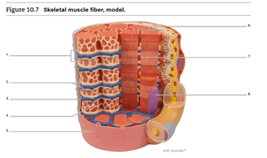
Skeletal Muscle Fiber Model
Skeletal muscle fibers are multinucleated cells containing specialized structures for contraction. The model below illustrates key components:
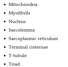
Mitochondria: Energy production
Myofibrils: Contractile units
Nucleus: Genetic control
Sarcolemma: Cell membrane
Sarcoplasmic reticulum: Calcium storage
Terminal cisternae: Enlarged SR regions
T-tubule: Transmits action potential
Triad: T-tubule + 2 terminal cisternae
Sarcomere Structure and Muscle Contraction
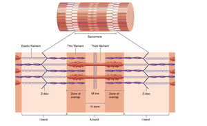
The sarcomere is the basic contractile unit of muscle, composed of thick and thin filaments. Muscle contraction occurs through the sliding filament mechanism.
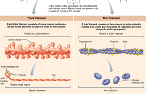
Thick filament: Myosin molecules with heads for binding actin
Thin filament: Actin, troponin, and tropomyosin
Z-disc: Boundary of sarcomere
M line: Center of sarcomere
H zone: Region with only thick filaments
I band: Region with only thin filaments
A band: Region with thick and thin filaments
Order of Muscle Contraction
Action potential arrives at neuromuscular junction
Acetylcholine released, depolarizes sarcolemma
Action potential travels down T-tubules
Calcium released from sarcoplasmic reticulum
Calcium binds to troponin, moves tropomyosin
Myosin binds to actin, power stroke occurs
ATP binds to myosin, detachment and reset
Isotonic vs. Isometric Contraction
Isotonic contraction: Muscle changes length, movement occurs (e.g., lifting weights)
Isometric contraction: Muscle length does not change, tension increases (e.g., holding a weight steady)
Muscles of the Body
Head, Neck, and Vertebrae Muscles
These muscles control facial expression, mastication, tongue movement, and neck posture.
Buccinator, masseter, temporalis: Chewing
Orbicularis oculi, orbicularis oris: Eye and mouth movement
Sternocleidomastoid, scalene: Neck movement
Frontalis, occipitalis: Forehead and scalp movement
Abdominal and Pelvic Muscles
These muscles support the trunk, aid in respiration, and control pelvic floor functions.
Rectus abdominis, external/internal oblique, transversus abdominis: Trunk flexion and rotation
Diaphragm: Main muscle of respiration
Levator ani, superficial transverse perineal: Pelvic floor support
Pectoral Girdle and Upper Limb Muscles
These muscles move the shoulder, arm, forearm, and hand.
Deltoid, pectoralis major/minor, trapezius: Shoulder movement
Biceps brachii, triceps brachii, brachialis: Arm flexion/extension
Flexor/extensor carpi, palmaris longus: Wrist movement
Pelvic Girdle and Lower Limb Muscles
These muscles control movement of the hip, thigh, leg, and foot.
Gluteus maximus/medius/minimus: Hip extension/abduction
Adductor group, gracilis, sartorius: Thigh movement
Quadriceps (rectus femoris, vastus group): Knee extension
Hamstrings (biceps femoris, semimembranosus, semitendinosus): Knee flexion
Gastrocnemius, soleus, tibialis anterior: Foot movement
Summary Table: Major Nerve Plexuses
The following tables summarize the main nerves, roots, and functions for each plexus:
Cervical Plexus Nerve | Nerve Roots | Structures Innervated |
|---|---|---|
Lesser occipital | C2 | Scalp and posterior ear |
Greater auricular | C2–C3 | Skin on ear |
Ansa cervicalis | C1–C3 | Geniohyoid and infrahyoid muscles |
Transverse cervical | C2–C3 | Skin on superior chest/shoulder |
Supraclavicular | C3–C4 | Skin on superior chest/shoulder |
Phrenic | C3–C5 | Diaphragm (motor) |
Brachial Plexus Nerve | Nerve Roots |
|---|---|
Axillary | C5–C6 |
Radial | C5–T1 |
Musculocutaneous | C5–C7 |
Median | C6–T1 |
Ulnar | C8–T1 |
Lumbar Plexus Nerve | Nerve Roots |
|---|---|
Iliohypogastric | L1 |
Ilioinguinal | L1 |
Genitofemoral | L1–L2 |
Femoral | L2–L4 |
Lateral femoral cutaneous | L2–L3 |
Obturator | L2–L4 |
Sacral Plexus Nerve | Nerve Roots | Structures Innervated |
|---|---|---|
Superior gluteal | L4–S1 | Gluteus medius/minimus, tensor fascia latae |
Inferior gluteal | L5–S2 | Gluteus maximus |
Pudendal | S2–S4 | Pelvic floor muscles |
Posterior femoral cutaneous | S1–S3 | Skin of posterior thigh |
Sciatic | L4–S3 | Leg (motor/sensory) |
Common fibular | L4–S2 | Leg/foot |
Tibial | L4–S3 | Leg/foot |
Key Muscle Fiber Structures and Functions
Mitochondria: Provide ATP for contraction
Myofibrils: Contain sarcomeres
Nucleus: Controls cell function
Sarcolemma: Muscle cell membrane
Sarcoplasmic reticulum: Stores calcium
Terminal cisternae: Release calcium
T-tubule: Conducts action potential
Triad: T-tubule flanked by terminal cisternae
Muscle Contraction Formula
The force generated by muscle contraction can be described by:
Where F is total force, n is the number of contracting fibers, and fsingle is the force per fiber.
Summary
This guide covers the peripheral nervous system, special senses, muscle tissue types, and the anatomy and physiology of muscle contraction, providing a comprehensive overview for ANP college students.
