Back
BackPeripheral Nervous System: Spinal Nerves, Plexuses, and Innervation
Study Guide - Smart Notes
 Tailored notes based on your materials, expanded with key definitions, examples, and context.
Spinal Nerves and Their Structure
Anatomy of Spinal Nerves
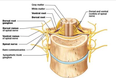
Spinal nerves are mixed nerves that emerge from the spinal cord and are responsible for transmitting motor, sensory, and autonomic signals between the spinal cord and the body. Each spinal nerve is formed by the union of a dorsal (sensory) root and a ventral (motor) root.
Dorsal root: Contains sensory (afferent) fibers and has a dorsal root ganglion, which houses the cell bodies of sensory neurons.
Ventral root: Contains motor (efferent) fibers.
Spinal nerve: The short segment formed by the union of dorsal and ventral roots; quickly branches into dorsal and ventral rami.
Dorsal ramus: Innervates intrinsic muscles of the back and dorsal skin.
Ventral ramus: Innervates lateral and anterior trunk, limbs, and forms nerve plexuses.
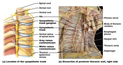
Rami communicantes: Connect spinal nerves to the sympathetic trunk ganglia, part of the autonomic nervous system.
Intercostal Nerves and Branches
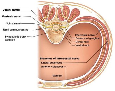
Ventral rami of thoracic spinal nerves (T2-T12) do not form plexuses but become intercostal nerves, which innervate the intercostal muscles and skin of the thorax.
Intercostal nerve: Provides motor and sensory innervation to the thoracic wall.
Branches: Lateral and anterior cutaneous branches supply the skin.
Nerve Plexuses
Cervical Plexus
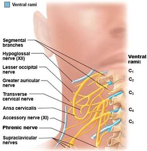
The cervical plexus is formed by ventral rami of C1-C4 (and part of C5), located in the neck. It provides sensory and motor innervation to the neck and upper shoulder.
Major nerves: Phrenic nerve (motor to diaphragm), lesser occipital, greater auricular, transverse cervical, supraclavicular nerves.
Accessory nerve (XI): Partly associated with the plexus.
Brachial Plexus
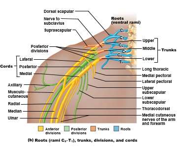
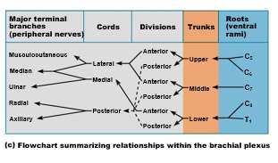
The brachial plexus is formed by ventral rami of C5-T1 and supplies the upper limb. It is organized into roots, trunks, divisions, cords, and terminal branches.
Roots: C5, C6, C7, C8, T1
Trunks: Upper, middle, lower
Divisions: Anterior and posterior
Cords: Lateral, medial, posterior
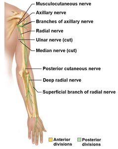
Major terminal branches: Musculocutaneous, median, ulnar, radial, axillary nerves
Innervation of the Upper Limb
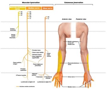
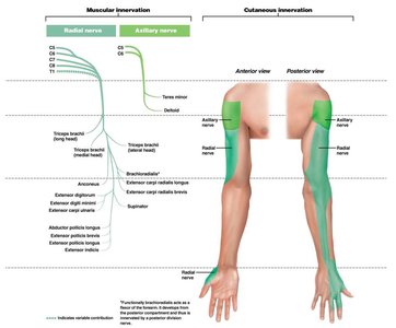
The terminal branches of the brachial plexus provide motor and sensory innervation to the muscles and skin of the arm and forearm.
Musculocutaneous nerve: Innervates anterior arm muscles and lateral forearm skin.
Median nerve: Innervates most forearm flexors and lateral hand skin.
Ulnar nerve: Innervates some forearm flexors and medial hand skin.
Radial nerve: Innervates posterior arm and forearm muscles and skin.
Axillary nerve: Innervates deltoid and teres minor muscles, skin over deltoid.
Lumbar Plexus
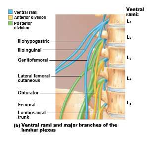
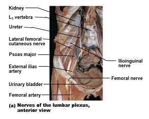
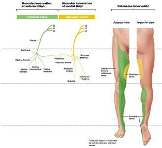
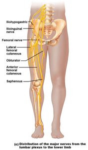
The lumbar plexus is formed by ventral rami of L1-L4 and lies within the psoas major muscle. It innervates the anterior and medial thigh, as well as parts of the perineum and lower abdomen.
Major nerves: Femoral nerve (anterior thigh), obturator nerve (medial thigh), lateral femoral cutaneous, iliohypogastric, ilioinguinal, genitofemoral nerves.
Sacral Plexus
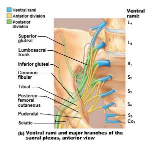
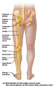
The sacral plexus is formed by ventral rami of L4-S4 and lies caudal to the lumbar plexus. It innervates the posterior thigh, most of the leg and foot, and parts of the pelvis.
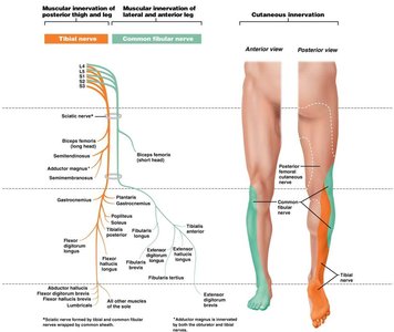
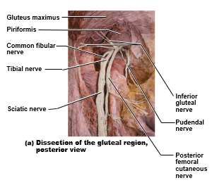
Sciatic nerve: Largest nerve, composed of tibial and common fibular nerves; innervates posterior thigh and leg.
Tibial nerve: Innervates posterior leg and plantar foot.
Common fibular nerve: Innervates lateral and anterior leg.
Other branches: Superior and inferior gluteal, pudendal, posterior femoral cutaneous nerves.
Dermatomes and Clinical Correlates
Dermatomes
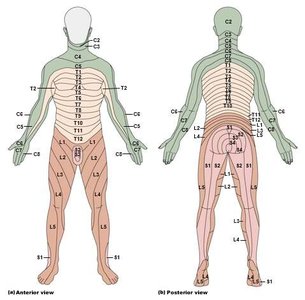
A dermatome is an area of skin innervated by the sensory fibers of a single spinal nerve. Dermatomes are important for diagnosing nerve injuries and viral infections such as shingles.
All spinal nerves except C1 have associated dermatomes.
Clinical application: Mapping dermatomes helps localize neurological lesions.
Shingles (Herpes Zoster)
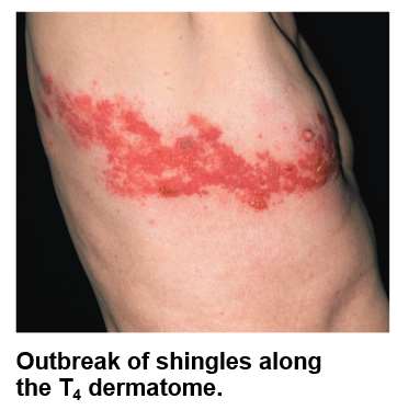
Shingles is a viral infection caused by the reactivation of the varicella-zoster virus, which remains dormant in the dorsal root ganglion after childhood chickenpox. It causes a painful rash along the affected dermatome.
Symptoms: Painful, blistering rash localized to one dermatome.
Pathophysiology: Virus travels along sensory nerve to skin.
Autonomic Nervous System (ANS) Overview
Divisions of the ANS
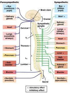
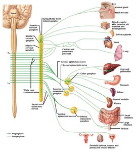
The autonomic nervous system (ANS) is the visceral motor division of the peripheral nervous system, controlling involuntary functions. It consists of two main divisions: sympathetic and parasympathetic.
Sympathetic division: Thoracolumbar origin; preganglionic neurons are short, ganglia near vertebral column.
Parasympathetic division: Craniosacral origin; preganglionic neurons are long, ganglia near or within target organs.
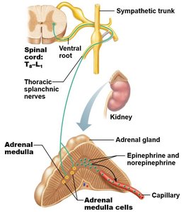
2-neuron pathway: Preganglionic neuron synapses with postganglionic neuron at autonomic ganglia.
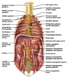
ANS Nerve Plexuses and Ganglia
Autonomic nerves pass through complex plexuses and ganglia to reach their target organs.
Sympathetic trunk ganglia: Paired ganglia along the vertebral column.
Collateral (prevertebral) ganglia: Unpaired, located on abdominal aorta in abdominopelvic cavity.
Parasympathetic plexuses: Vagus nerve and pelvic splanchnic nerves pass through various plexuses.
Special Senses: The Eye
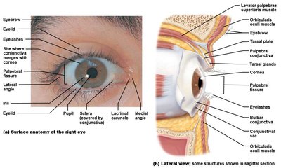
External and Accessory Structures
The eye is protected and moved by several external structures, including the eyelids, conjunctiva, and extrinsic muscles.
Protection: Eyebrows, eyelids, conjunctiva, lacrimal apparatus.
Movement: Four rectus and two oblique muscles per eye.
Internal Anatomy of the Eye
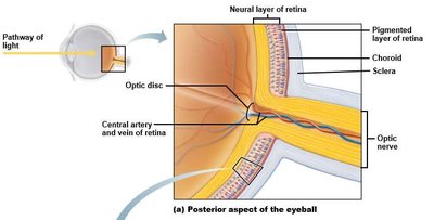
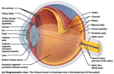
The eye is divided into anterior and posterior segments by the lens, and its wall consists of three layers: fibrous, vascular, and sensory tunics.
Fibrous tunic: Sclera and cornea.
Vascular tunic: Choroid, ciliary body, iris.
Sensory tunic: Retina and optic nerve.
Retina specializations: Macula lutea, fovea centralis, optic disc.