 Back
BackPeripheral Nervous System: Structure, Classification, and Major Nerve Plexuses
Study Guide - Smart Notes
 Tailored notes based on your materials, expanded with key definitions, examples, and context.
Peripheral Nervous System (PNS) Overview
Structural Organization of the Nervous System
The nervous system is divided into the central nervous system (CNS) and the peripheral nervous system (PNS). The PNS includes all neural structures outside the brain and spinal cord, such as sensory receptors, sensory and motor neurons, and nerve endings where motor neurons synapse with muscle or gland cells.
Sensory (afferent) division: Transmits sensory information to the CNS.
Motor (efferent) division: Transmits commands from the CNS to muscles and glands.
Somatic nervous system: Controls voluntary movements.
Autonomic nervous system (ANS): Controls involuntary functions and is subdivided into sympathetic and parasympathetic divisions.
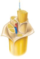
Nerves: Structure and Classification
Structure of a Nerve
A nerve is a cordlike organ of the PNS, consisting of bundles of myelinated and nonmyelinated peripheral axons enclosed by connective tissue. Nerves are classified as spinal or cranial depending on their origin.
Endoneurium: Loose connective tissue that encloses individual axons and their myelin sheaths.
Perineurium: Coarse connective tissue that bundles fibers into fascicles.
Epineurium: Tough fibrous sheath around all fascicles to form the nerve.
Tip: From outermost to innermost: Epineurium → Perineurium → Endoneurium.
Classification of Nerves
Mixed nerves: Contain both sensory and motor fibers; impulses travel both to and from the CNS.
Sensory (afferent) nerves: Carry impulses only toward the CNS.
Motor (efferent) nerves: Carry impulses only away from the CNS.
Most nerves are mixtures of afferent and efferent fibers, and somatic (voluntary) and autonomic (involuntary) fibers.
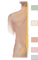
Spinal Nerves
Overview and Classification
There are 31 pairs of spinal nerves, all of which are mixed nerves. They supply all body parts except the head and part of the neck.
8 pairs of cervical nerves (C1–C8)
12 pairs of thoracic nerves (T1–T12)
5 pairs of lumbar nerves (L1–L5)
5 pairs of sacral nerves (S1–S5)
1 pair of coccygeal nerves (Co1)
Spinal Nerve Roots and Branches
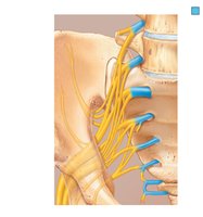
Each spinal nerve is connected to the spinal cord via two roots:
Ventral roots: Contain motor (efferent) fibers from ventral horn motor neurons that innervate skeletal muscles.
Dorsal roots: Contain sensory (afferent) fibers from sensory neurons in dorsal root ganglia that conduct impulses from peripheral receptors.
Both ventral and dorsal roots are branched medially as rootlets that join laterally to form the spinal nerve. Spinal roots are unmixed (carry only sensory or only motor neurons), but the roots mingle to form mixed spinal nerves, which exit the vertebral column via intervertebral foramina.
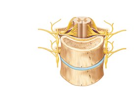
Rami of Spinal Nerves
After exiting the vertebral column, spinal nerves divide into branches called rami:
Dorsal ramus: Innervates the posterior trunk (skin and muscles of the back).
Ventral ramus: Innervates the lateral and anterior trunk, and the limbs.
Both dorsal and ventral rami are mixed nerves (carry both sensory and motor fibers).
Plexuses of the Peripheral Nervous System
Definition and Function
A plexus is a network of nerves that primarily serves the limbs. Within a plexus, fibers from several ventral rami crisscross and become redistributed, so each branch contains fibers from several spinal nerves. This arrangement ensures that damage to a single spinal nerve does not completely paralyze any limb muscle.
Cervical Plexus
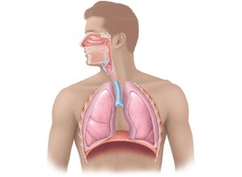
The cervical plexus is formed by ventral rami of C1–C4. It innervates the skin of the neck, ear, back of the head, and shoulders. The most important nerve is the phrenic nerve, which innervates the diaphragm and is essential for breathing.
Phrenic nerve: Arises from C3–C5; irritation causes hiccups; damage can cause respiratory arrest.
Brachial Plexus
The brachial plexus is formed by ventral rami of C5–C8 and T1. It gives rise to major nerves that innervate the upper limb, including the median, ulnar, and radial nerves.
Median nerve: Innervates flexor muscles in the forearm and hand; damage causes loss of pincer grasp and is associated with carpal tunnel syndrome.
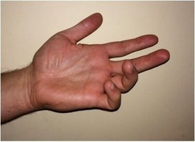
Ulnar nerve: Innervates forearm flexors and fingers 4–5; damage can cause "claw hand." The "funny bone" sensation is due to the ulnar nerve.
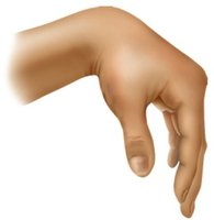
Radial nerve: Innervates triceps brachii and extensor muscles of the forearm and hand; damage results in "wrist drop."
Lumbosacral Plexus
The lumbosacral plexus consists of the lumbar and sacral plexuses, which have significant overlap. They serve the lower limb, abdomen, pelvis, and buttocks.
Lumbar plexus (L1–L4): Innervates thigh and abdominal wall; the femoral nerve innervates quadriceps femoris for thigh flexion and leg extension.
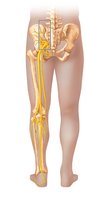
Sacral plexus (L4–S4): Innervates buttock, lower limb, and pelvis; the sciatic nerve is the largest nerve, innervating hamstrings and most muscles in the leg and foot.
Clinical Application: Sciatic Nerve and Sciatica
Damage to the proximal part of the sciatic nerve can result from herniated discs or improper injections in the buttocks. Sciatica is characterized by stabbing pain radiating along the sciatic nerve. Severe injury can cause paralysis of the hamstrings and muscles below the knee, resulting in "footdrop."
Summary Table: Major Spinal Nerves and Plexuses
Region | Spinal Nerves | Plexus | Major Nerves | Key Muscles/Areas Innervated |
|---|---|---|---|---|
Cervical | C1–C8 | Cervical plexus | Phrenic nerve | Neck, diaphragm |
Brachial | C5–T1 | Brachial plexus | Median, Ulnar, Radial nerves | Upper limb |
Lumbar | L1–L4 | Lumbar plexus | Femoral nerve | Thigh, abdominal wall |
Sacral | L4–S4 | Sacral plexus | Sciatic nerve | Buttock, lower limb |
Key Concepts and Clinical Correlations
Nerve regeneration: PNS nerves can regenerate if the soma is intact and damage is not severe; CNS nerves do not regenerate.
Plexus injury: Damage to a single spinal nerve does not cause complete paralysis due to overlapping innervation.
Clinical syndromes: Carpal tunnel syndrome (median nerve), claw hand (ulnar nerve), wrist drop (radial nerve), and sciatica (sciatic nerve).
