 Back
BackPeripheral Nervous System: Structure, Cranial Nerves, and Sensory Receptors
Study Guide - Smart Notes
 Tailored notes based on your materials, expanded with key definitions, examples, and context.
Peripheral Nervous System (PNS) Overview
Structure of a Nerve
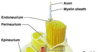
The peripheral nervous system consists of nerves that connect the central nervous system (CNS) to the rest of the body. Each nerve is a bundle of axons, blood vessels, and connective tissue layers that provide structural support and protection.
Axon: The long, slender projection of a neuron that conducts electrical impulses away from the cell body.
Myelin sheath: An insulating layer around many axons, increasing the speed of nerve impulse transmission.
Endoneurium: A delicate connective tissue surrounding individual axons.
Perineurium: A connective tissue sheath that groups axons into bundles called fascicles.
Epineurium: The outermost connective tissue layer that encloses the entire nerve, including multiple fascicles and blood vessels.
Fascicle: A bundle of axons within a nerve.
Blood vessels: Supply nutrients and oxygen to nerve tissues.
Cranial Nerves
Classification and Function
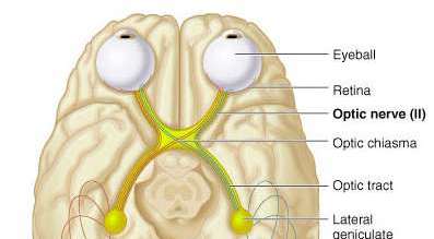
Cranial nerves are twelve pairs of nerves that emerge directly from the brain. Each has specific sensory, motor, or mixed functions. Some also carry parasympathetic fibers.
Cranial Nerve | Sensory Function | Motor Function | Parasympathetic (PS) Fibers |
|---|---|---|---|
I Olfactory | Yes (smell) | No | No |
II Optic | Yes (vision) | No | No |
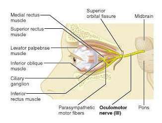
III Oculomotor | No | Yes | Yes |
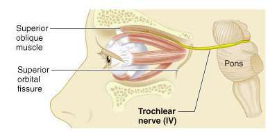
IV Trochlear | No | Yes | No |
V Trigeminal | Yes (general sensation) | Yes | No |
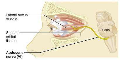
VI Abducens | No | Yes | No |
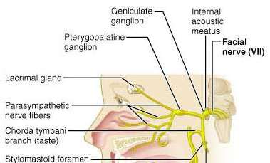
VII Facial | Yes (taste) | Yes | Yes |
VIII Vestibulocochlear | Yes (hearing and balance) | No | No |
IX Glossopharyngeal | Yes (taste) | Yes | Yes |
X Vagus | Yes (taste) | Yes | Yes |
XI Accessory | No | Yes | No |
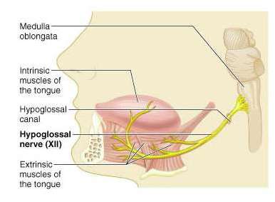
XII Hypoglossal | No | Yes | No |
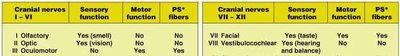
PS = parasympathetic
Selected Cranial Nerves: Structure and Pathways
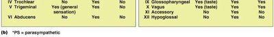
Optic Nerve (II): Transmits visual information from the retina to the brain. The optic chiasma is where fibers partially cross, allowing visual fields to be processed by both hemispheres.
Oculomotor Nerve (III): Controls most of the eye's movements, the constriction of the pupil, and maintains an open eyelid.
Trochlear Nerve (IV): Innervates the superior oblique muscle, enabling downward and lateral eye movement.
Abducens Nerve (VI): Controls the lateral rectus muscle, responsible for abducting the eye (moving it laterally).
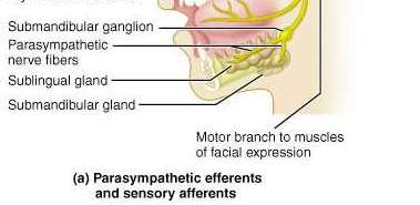
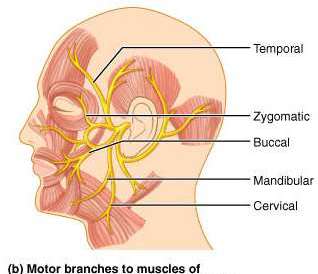
Facial Nerve (VII): Provides motor innervation to facial expression muscles and carries taste sensations from the anterior two-thirds of the tongue. It also carries parasympathetic fibers to salivary and lacrimal glands.
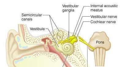
Vestibulocochlear Nerve (VIII): Responsible for hearing and balance, with vestibular and cochlear branches.
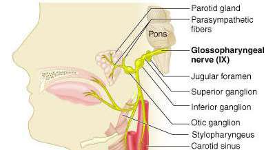
Glossopharyngeal Nerve (IX): Involved in taste, swallowing, and salivation; carries parasympathetic fibers to the parotid gland.
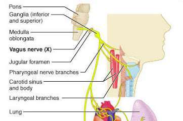
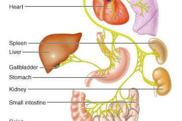
Vagus Nerve (X): The only cranial nerve to extend beyond the head and neck, innervating thoracic and abdominal organs. It is crucial for parasympathetic control of the heart, lungs, and digestive tract.
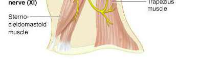
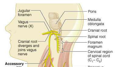
Accessory Nerve (XI): Supplies the sternocleidomastoid and trapezius muscles, aiding in head movement.
Hypoglossal Nerve (XII): Controls tongue movements essential for speech and swallowing.
Spinal Nerves and Plexuses
Spinal Nerve Organization
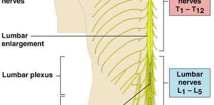
Spinal nerves emerge from the spinal cord and are organized into regions: cervical, thoracic, lumbar, sacral, and coccygeal. Each region forms nerve plexuses that innervate specific body areas.
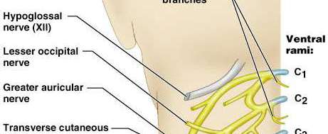
Cervical plexus: Innervates the neck and diaphragm.
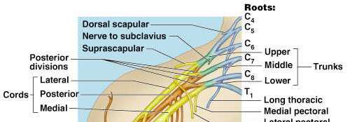
Brachial plexus: Supplies the shoulders and upper limbs.
Lumbar plexus: Innervates the lower abdomen, anterior and medial thigh.
Sacral plexus: Supplies the pelvis, posterior thigh, and most of the lower limb.
Cauda equina: A bundle of spinal nerves at the lower end of the spinal cord.
Cervical and Brachial Plexuses
Cervical plexus: Contains nerves such as the phrenic nerve, which controls the diaphragm.
Brachial plexus: Gives rise to nerves of the upper limb, including the musculocutaneous, median, ulnar, radial, and axillary nerves.
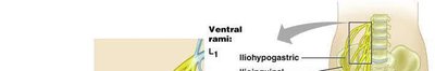
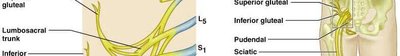
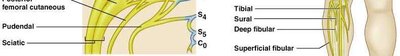
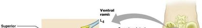
Lumbar and Sacral Plexuses
Lumbar plexus: Includes the femoral and obturator nerves, serving the anterior and medial thigh.
Sacral plexus: Contains the sciatic nerve, the largest nerve in the body, serving the lower limb.
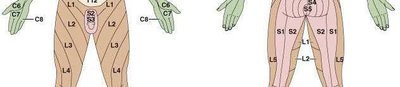
Dermatomes
A dermatome is an area of skin supplied by sensory fibers from a single spinal nerve. Mapping dermatomes helps diagnose nerve or spinal cord injuries.
General Sensory Receptors
Classification by Structure and Function
Sensory receptors detect changes in the environment and transmit information to the CNS. They are classified by structure (encapsulated or unencapsulated) and function (type of stimulus detected).
Structural Class | Illustration | Functional Class (Location/Stimulus) | Body Location |
|---|---|---|---|
Unencapsulated (Free nerve endings) | — | L: Exteroceptors, interoceptors, proprioceptors S: Nociceptors (pain), thermoreceptors (temp), possible mechanoreceptors | Most body tissues, especially connective tissues and epithelia |
Merkel discs (modified free nerve endings) | — | L: Exteroceptors S: Mechanoreceptors (light pressure, slowly adapting) | Basal layer of epidermis of skin |
Meissner's corpuscles | — | L: Exteroceptors S: Mechanoreceptors (light pressure, discriminative touch, vibration of low frequency, rapidly adapting) | Dermal papillae of hairless skin (fingertips, soles, eyelids, etc.) |
Pacini corpuscles | — | L: Exteroceptors, interoceptors, some proprioceptors S: Mechanoreceptors (deep pressure, stretch, vibration of high frequency, rapidly adapting) | Subcutaneous tissue, periostea, tendons, ligaments, joint capsules |
Ruffini corpuscles | — | L: Exteroceptors, proprioceptors S: Mechanoreceptors (deep pressure, stretch, slowly or nonadapting) | Deep in dermis, hypodermis, joint capsules |
Muscle spindles | — | L: Proprioceptors S: Mechanoreceptors (muscle stretch) | Skeletal muscles, especially extremities |
Golgi tendon organs | — | L: Proprioceptors S: Mechanoreceptors (tendon stretch) | Tendons |
Joint kinesthetic receptors | — | L: Proprioceptors S: Mechanoreceptors and nociceptors | Joint capsules of synovial joints |
Reflex Arcs
Components of a Reflex Arc
A reflex arc is the basic functional unit of the nervous system, allowing for rapid, automatic responses to stimuli. It consists of five main components:
Receptor: Detects the stimulus.
Sensory neuron: Transmits afferent impulses to the CNS.
Integration center: Processes information (may involve interneurons).
Motor neuron: Conducts efferent impulses from the integration center to an effector.
Effector: Muscle or gland that responds to the impulse.
Somatic vs. Autonomic Nervous System
Comparison of Pathways and Effectors
The somatic and autonomic nervous systems differ in their effectors, pathways, and neurotransmitters.
Somatic nervous system: Controls voluntary movements via a single neuron pathway to skeletal muscle, using acetylcholine as the neurotransmitter.
Autonomic nervous system: Controls involuntary functions (smooth muscle, cardiac muscle, glands) via a two-neuron chain (preganglionic and postganglionic neurons). The sympathetic division uses norepinephrine, while the parasympathetic division uses acetylcholine at the effector.
Additional info: The notes above integrate and expand upon the provided images and tables, ensuring a comprehensive overview of the peripheral nervous system, cranial nerves, sensory receptors, and reflexes, as relevant to an introductory college-level anatomy and physiology course.
