 Back
Backch. 13 part A; Peripheral Nervous System: Structure, Function, and Sensory Processing
Study Guide - Smart Notes
 Tailored notes based on your materials, expanded with key definitions, examples, and context.
The Peripheral Nervous System (PNS)
Overview and Functional Organization
The Peripheral Nervous System (PNS) connects the central nervous system (CNS) to the external and internal environments, enabling communication between the brain, spinal cord, and the rest of the body. It consists of all neural structures outside the brain and spinal cord and is divided into sensory and motor divisions, with further subdivisions for somatic and autonomic functions.
Sensory (Afferent) Division: Transmits sensory information from receptors to the CNS.
Motor (Efferent) Division: Carries motor commands from the CNS to effectors (muscles and glands).
Somatic Nervous System: Controls voluntary movements of skeletal muscles.
Autonomic Nervous System (ANS): Regulates involuntary functions (e.g., heart rate, digestion) and is further divided into sympathetic and parasympathetic divisions.
Sensory Receptors
Definition and Function
Sensory receptors are specialized structures that detect changes (stimuli) in the environment. Their activation generates graded potentials, which may trigger nerve impulses sent to the CNS for processing. Sensation is the awareness of a stimulus, while perception is the interpretation of its meaning.
Classification of Sensory Receptors
By Stimulus Type
Mechanoreceptors: Respond to touch, pressure, vibration, and stretch.
Thermoreceptors: Detect temperature changes.
Photoreceptors: Respond to light (e.g., in the retina).
Chemoreceptors: Detect chemicals (e.g., smell, taste, blood chemistry).
Nociceptors: Respond to potentially damaging stimuli (pain).
By Location
Exteroceptors: Detect stimuli from outside the body (e.g., skin, special sense organs).
Interoceptors (Visceroceptors): Monitor internal environment (e.g., viscera, blood vessels).
Proprioceptors: Sense body position and movement (e.g., muscles, tendons, joints).
By Structural Complexity
Simple Receptors (General Senses): Modified dendritic endings of sensory neurons; found throughout the body.
Complex Receptors (Special Senses): Located in sense organs for vision, hearing, equilibrium, smell, and taste.
Structural Types of General Sensory Receptors
Nonencapsulated (Free) Nerve Endings
Primarily detect temperature, pain, and light touch.
Include thermoreceptors, nociceptors, tactile (Merkel) discs, and hair follicle receptors.
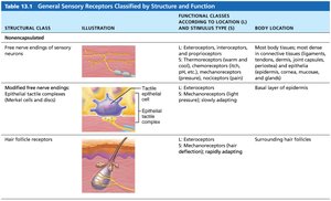
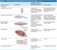
Encapsulated Dendritic Endings
Mechanoreceptors with terminal endings encased in connective tissue.
Include tactile (Meissner's) corpuscles, lamellar (Pacinian) corpuscles, bulbous (Ruffini) endings, muscle spindles, tendon organs, and joint kinesthetic receptors.
Sensory Processing
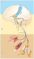
Somatosensory System
The somatosensory system processes sensory input from the body wall and limbs. It receives information from exteroceptors, proprioceptors, and interoceptors, relaying it toward the brain and processing it at multiple levels.
Levels of Neural Integration
Receptor Level: Sensory reception and transmission to the CNS.
Circuit Level: Processing in ascending pathways to the brain.
Perceptual Level: Processing in cortical sensory areas for conscious perception.
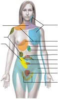
Perception of Pain
Visceral Pain: Originates from internal organs; often vague and poorly localized.
Referred Pain: Pain perceived at a location other than the site of the stimulus, due to convergence of visceral and somatic sensory fibers on the same pathways.
Example: Pain in the left arm during a heart attack.
Nerves and Associated Ganglia
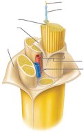
Structure of a Nerve
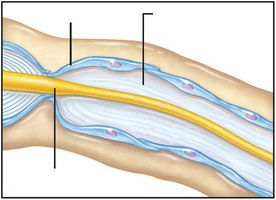
A nerve is a cordlike organ composed of bundles of axons (myelinated and unmyelinated) surrounded by connective tissue layers:
Endoneurium: Surrounds individual axons and their myelin sheaths.
Perineurium: Bundles groups of axons into fascicles.
Epineurium: Encloses all fascicles to form the nerve.
Classification of Nerves
Mixed Nerves: Contain both sensory (afferent) and motor (efferent) fibers; most common type.
Sensory (Afferent) Nerves: Carry impulses toward the CNS.
Motor (Efferent) Nerves: Carry impulses away from the CNS.
Mixed nerves may contain somatic and autonomic fibers.
Ganglia
Ganglia: Collections of neuron cell bodies in the PNS.
Sensory Ganglia: Dorsal root ganglia (contain sensory neuron cell bodies).
Autonomic Ganglia: Contain cell bodies of autonomic motor neurons.
Regeneration of Nerve Fibers
Regeneration in the CNS
Most CNS axons do not regenerate due to inhibitory proteins from oligodendrocytes and scar tissue formation by astrocytes.
Current research focuses on overcoming these barriers to promote regeneration.
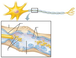
Regeneration in the PNS
PNS axons can regenerate if the cell body is intact and damage is not severe. The process involves several steps:
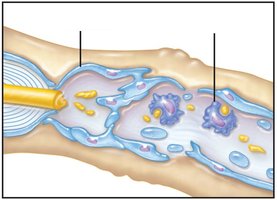
Wallerian Degeneration: The axon and myelin sheath distal to the injury degenerate.
Cleanup: Macrophages and Schwann cells remove debris and stimulate Schwann cell proliferation.
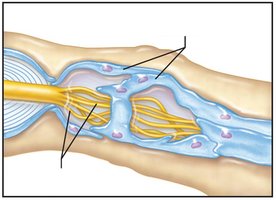
Regeneration Tube Formation: Schwann cells align to form a tube guiding new axon sprouts.
Axon Regrowth and Remyelination: The axon regenerates and is remyelinated by Schwann cells.
Example: Peripheral nerve injuries, such as those from trauma, may recover function if the axon regenerates successfully through these steps.
Additional info: CNS regeneration is a major focus of neuroscience research due to its implications for spinal cord injury and neurodegenerative diseases.
