 Back
BackPns 1
Study Guide - Smart Notes
 Tailored notes based on your materials, expanded with key definitions, examples, and context.
Peripheral Nervous System (PNS)
Overview of the Peripheral Nervous System
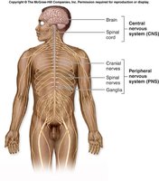
The Peripheral Nervous System (PNS) consists of all nervous structures outside the brain and spinal cord. It serves as the communication lines that link all parts of the body to the central nervous system (CNS). The PNS includes nerves, ganglia, and sensory receptors.
Nerves: Bundles of axons (nerve fibers) outside the CNS.
Ganglia: Collections of neuron cell bodies in the PNS.
Sensory receptors: Specialized structures that detect changes in the environment.
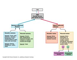
Functional Organization of the PNS
The PNS is functionally divided into sensory (afferent) and motor (efferent) divisions. Sensory information flows toward the CNS, while motor commands flow away from the CNS to effector organs.
Sensory (Afferent) Division: Transmits impulses from sensory receptors to the CNS.
Motor (Efferent) Division: Transmits impulses from the CNS to effector organs (muscles and glands).
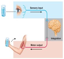
Functions of the Peripheral Nervous System
The PNS is responsible for relaying sensory information to the CNS for processing and carrying out motor commands from the CNS to the body.
Sensory Input: Detects stimuli and sends information to the CNS.
Motor Output: Executes responses by activating muscles or glands.
Nerves and Ganglia
Definitions and Terminology
Understanding the terminology is crucial for distinguishing between different components of the nervous system:
Neuron: A nerve cell, the basic functional unit of the nervous system.
Nerve Fiber: A long axon of a neuron.
Nerve: A bundle of nerve fibers (axons) in the PNS.
Ganglion (plural: ganglia): A cluster of neuron cell bodies in the PNS.
Nerves can be composed of myelinated or unmyelinated axons.
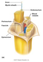
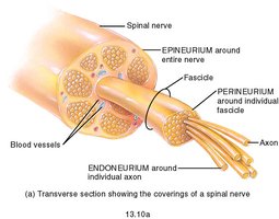
Nerve Anatomy
Nerves are organized into bundles called fascicles, which are separated by connective tissue layers:
Endoneurium: Surrounds individual axons.
Perineurium: Surrounds each fascicle (bundle of axons).
Epineurium: Encloses the entire nerve.
Classification of Nerves
Functional Classification
Nerves are classified by the direction in which they transmit action potentials relative to the CNS:
Sensory (Afferent) Nerves: Carry information toward the CNS.
Motor (Efferent) Nerves: Carry information away from the CNS.
Mixed Nerves: Contain both sensory and motor fibers; transmit information in both directions.
Classification by Location
Nerves are also classified based on their attachment to the CNS:
Cranial Nerves: Attach to the brain.
Spinal Nerves: Attach to the spinal cord.
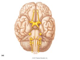
Cranial Nerves
Overview of Cranial Nerves
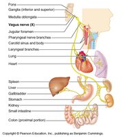
There are 12 pairs of cranial nerves, each identified by a name, Roman numeral, location of attachment, function, and functional classification (sensory, motor, or mixed). These nerves primarily serve the head and neck, with the exception of the vagus nerve, which extends into the thorax and abdomen.
Number | Name | Attachment | Function | Classification |
|---|---|---|---|---|
I | Olfactory | Cerebrum | Smell | Sensory |
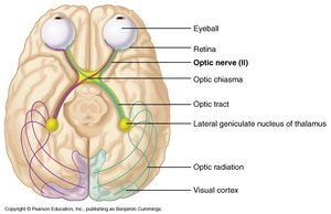
II | Optic | Thalamus | Vision | Sensory |
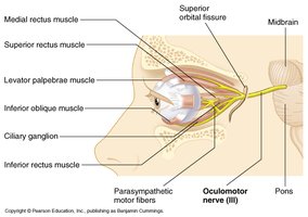
III | Oculomotor | Midbrain | Eye movement, pupil constriction | Motor |
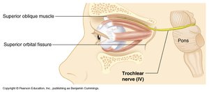
IV | Trochlear | Midbrain | Eye movement (superior oblique) | Motor |
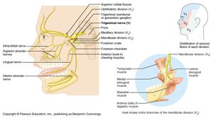
V | Trigeminal | Pons | Facial sensation, mastication | Mixed |
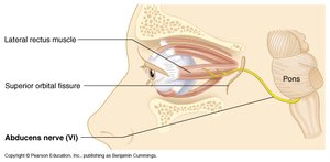
VI | Abducens | Pons | Eye movement (lateral rectus) | Motor |
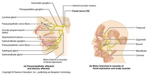
VII | Facial | Pons | Facial expression, taste (anterior 2/3 tongue) | Mixed |
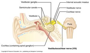
VIII | Vestibulocochlear | Medulla | Hearing, balance | Sensory |
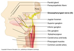
IX | Glossopharyngeal | Medulla | Swallowing, taste (posterior 1/3 tongue) | Mixed |
X | Vagus | Medulla | Viscera, heart, lungs, GI tract | Mixed |
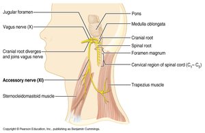
XI | Accessory | Medulla | Neck muscles, shoulder shrug | Motor |
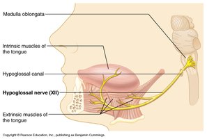
XII | Hypoglossal | Medulla | Tongue movement | Motor |
Selected Cranial Nerves and Their Functions
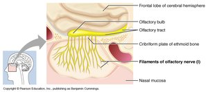
Olfactory (I): Smell; sensory only.
Optic (II): Vision; sensory only.
Oculomotor (III): Eye movement, pupil constriction; motor only.
Trochlear (IV): Superior oblique muscle of eye; motor only.
Trigeminal (V): Facial sensation, chewing; mixed.
Abducens (VI): Lateral rectus muscle of eye; motor only.
Facial (VII): Facial expression, taste; mixed.
Vestibulocochlear (VIII): Hearing and balance; sensory only.
Glossopharyngeal (IX): Swallowing, taste; mixed.
Vagus (X): Parasympathetic control of heart, lungs, GI tract; mixed.
Accessory (XI): Neck muscles; motor only.
Hypoglossal (XII): Tongue movement; motor only.
Mnemonic Devices for Cranial Nerves
Name Order: "Oh, Oh, Oh, To Touch And Feel Very Good Velvet, Ah Ha!" (First letter of each word = first letter of each nerve in order)
Functional Classification: "Some Say Marry Money, But My Brother Says Big Brains Matter More" (S = sensory, M = motor, B = both/mixed)
Spinal Nerves
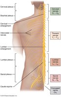
Overview of Spinal Nerves
There are 31 pairs of spinal nerves, all classified as mixed nerves (containing both sensory and motor fibers). They are named according to the region of the vertebral column from which they emerge:
8 pairs of cervical nerves (C1–C8)
12 pairs of thoracic nerves (T1–T12)
5 pairs of lumbar nerves (L1–L5)
5 pairs of sacral nerves (S1–S5)
1 pair of coccygeal nerves (C0)

Spinal Nerve Structure
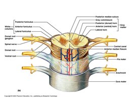
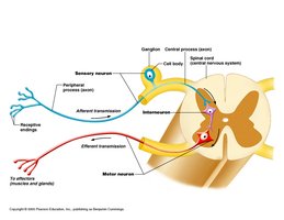
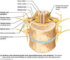
Each spinal nerve is formed by the union of a dorsal (sensory) root and a ventral (motor) root. The dorsal root contains sensory fibers whose cell bodies are in the dorsal root ganglion, while the ventral root contains motor fibers whose cell bodies are in the anterior horn of the spinal cord.

Rami and Plexuses
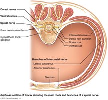
After emerging from the spinal cord, each spinal nerve splits into dorsal and ventral rami. The rami communicantes connect to the sympathetic trunk ganglia, part of the autonomic nervous system.
Dorsal Ramus: Innervates the posterior trunk or neck.
Ventral Ramus: Innervates anterior and lateral trunk and limbs.

Spinal Nerve Plexuses
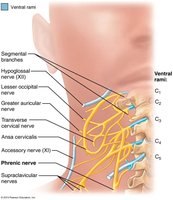
Cervical Plexus
The cervical plexus is formed by the ventral rami of C1–C4. Its most important nerve is the phrenic nerve (C3–C5), which innervates the diaphragm and is essential for breathing.
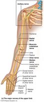
Brachial Plexus
The brachial plexus is formed by the ventral rami of C5–C8 and T1. It gives rise to nerves that innervate the upper limb, including:
Axillary (C5, C6)
Radial (C5–C8, T1)
Median (C5–C8, T1)
Musculocutaneous (C5–C7)
Ulnar (C8, T1)
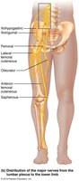
Lumbar Plexus
The lumbar plexus is formed by the ventral rami of L1–L4. Major nerves include:
Femoral (L2–L4)
Obturator (L2–L4)
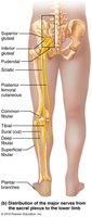
Sacral Plexus
The sacral plexus is formed by the ventral rami of L4–S4. Its most important nerve is the sciatic nerve (L4–S4), the longest nerve in the body, which divides into the tibial and common fibular nerves.
Summary Table: Major Spinal Nerve Plexuses and Key Nerves
Plexus | Spinal Nerves | Key Nerves | Major Regions Served |
|---|---|---|---|
Cervical | C1–C4 | Phrenic | Neck, diaphragm |
Brachial | C5–T1 | Axillary, Radial, Median, Musculocutaneous, Ulnar | Shoulder, arm, hand |
Lumbar | L1–L4 | Femoral, Obturator | Anterior thigh, medial thigh |
Sacral | L4–S4 | Sciatic (tibial & common fibular) | Posterior thigh, lower leg, foot |
Additional info: The thoracic nerves (T2–T12) do not form a plexus and are known as intercostal nerves, which innervate the intercostal muscles and skin of the thorax.
