 Back
Peripheral Nervous System: Structure, Function, and Clinical Relevance
Study Guide - Smart Notes
 Tailored notes based on your materials, expanded with key definitions, examples, and context.
Peripheral Nervous System (PNS)
Overview and Functional Divisions
The Peripheral Nervous System (PNS) connects the Central Nervous System (CNS) to the rest of the body, enabling sensory input and motor output. It is functionally divided into sensory (afferent) and motor (efferent) divisions, each further subdivided into somatic and visceral components, as well as general and special senses.
Sensory Division: Transmits sensory information from receptors to the CNS.
Motor Division: Sends motor commands from the CNS to effectors (muscles and glands).
Somatic: Relates to body wall, limbs, and voluntary control.
Visceral: Relates to internal organs and involuntary control.
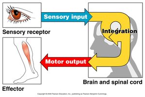
Organization of the PNS
The PNS consists of sensory receptors, nerves, ganglia, and motor endings.
Sensory Receptors: Detect stimuli from inside or outside the body.
Nerves: Bundles of peripheral axons transmitting signals.
Ganglia: Clusters of peripheral neuronal cell bodies.
Motor Endings: Axon terminals of motor neurons that innervate effectors.
Sensory Receptors
Types and Functions
Sensory receptors initiate signals in sensory axons and are classified based on their structure and function.
Free Nerve Endings: Monitor general sensory information such as pain and temperature.
Encapsulated Nerve Endings: Specialized for detecting pressure, pain, and stretch.
Complete Receptor Cells: Specialized epithelial cells or small neurons monitoring special sensory information (e.g., taste, vision).
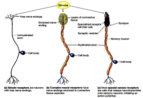
Examples of Sensory Receptors
General Sensory Receptors: Touch, pain, pressure, vibration, temperature, proprioception.
Special Sensory Receptors: Hearing, equilibrium, vision, taste, smell.
Proprioceptors
Types and Functions
Proprioceptors are specialized sensory receptors that monitor the position and movement of the body. There are three main types:
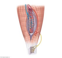
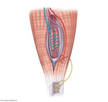
Muscle Spindles: Measure the changing length of a muscle and are embedded within the perimysium between muscle fascicles.
Tendon Organs (Golgi Tendon Organs): Located near the muscle-tendon junction, monitor tension within tendons.
Joint Kinesthetic Receptors: Sensory nerve endings within joint capsules, including lamellar corpuscles, bulbous corpuscles, free nerve endings, and receptors resembling tendon organs.
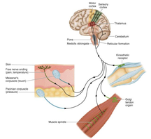
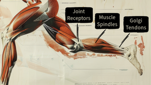
Muscle Spindles
Muscle spindles contain intrafusal muscle fibers and are sensitive to the rate and degree of muscle stretch.
Intrafusal Muscle Fibers: Modified skeletal muscle fibers within the spindle.
Annulospiral Endings: Primary sensory endings stimulated by stretch.
Flower Spray Endings: Secondary sensory endings.
Tendon Organs and Joint Kinesthetic Receptors
Tendon Organs: Monitor tension in tendons, preventing muscle damage from excessive force.
Joint Kinesthetic Receptors: Provide information about joint position and movement.
Cranial Nerves
Structure and Function
Cranial nerves are twelve pairs of nerves that attach to the brain and pass through specific foramina of the skull.
Numbered I to XII: Each has a specific function, primarily serving head and neck structures.
Vagus Nerve (X): The only cranial nerve extending into the abdomen.
Attachment: Cranial nerves I and II attach to the forebrain; others attach to the brain stem.
Spinal Nerves
Structure and Distribution
Spinal nerves connect the CNS to the body and are named for their point of issue from the spinal cord.
31 Pairs: 8 cervical, 12 thoracic, 5 lumbar, 5 sacral, 1 coccygeal.
Connection: Each spinal nerve connects to the spinal cord via dorsal (sensory) and ventral (motor) roots.
Branching: Spinal nerves branch into dorsal and ventral rami, which contain both sensory and motor fibers.
Rami Communicantes: Connect to the base of the ventral ramus and lead to the sympathetic chain ganglia.
Nerve Plexuses
Introduction and Major Plexuses
Nerve plexuses are networks formed by ventral rami (except T2–T12) that branch and join with one another, primarily serving the limbs.
Cervical Plexus: Formed by C1–C4, innervates neck muscles and skin; phrenic nerve innervates the diaphragm.
Brachial Plexus: Formed by C5–T1, innervates the upper limb; consists of roots, trunks, divisions, and cords.
Lumbar Plexus: Formed by L1–L4, innervates anterior thigh muscles (femoral nerve) and adductor muscles (obturator nerve).
Sacral Plexus: Formed by L4–S4, innervates the lower limb; includes the sciatic nerve.
Disorders of the PNS
Common Disorders
Shingles (Herpes Zoster): Viral infection stemming from childhood chicken pox, often triggered by stress and affecting people over age 50.
Migraine Headache: Related to sensory innervation of cerebral arteries; dilation compresses and irritates sensory nerve endings.
Peripheral Neuropathy: Pathological condition of peripheral nerves, causing paresthesia, pain, burning, loss of sensation, muscle weakness, and paralysis. Causes include trauma, repetitive use, HIV, diabetes, and vitamin B deficiency.
Summary Table: Functional Organization of the PNS
Division | Sensory Function | Motor Function |
|---|---|---|
Somatic | Touch, pain, pressure, vibration, temperature, proprioception | Motor innervation of skeletal muscles |
Visceral | Stretch, pain, temperature, chemical changes, irritation in viscera; nausea, hunger | Motor innervation of smooth muscle, cardiac muscle, glands |
Special | Hearing, equilibrium, vision, taste, smell | None |
Key Terms and Definitions
Peripheral Nervous System (PNS): The part of the nervous system outside the brain and spinal cord.
Central Nervous System (CNS): Composed of the brain and spinal cord.
Ganglia: Clusters of neuronal cell bodies in the PNS.
Effector: Muscle or gland that responds to motor output.
Proprioceptor: Sensory receptor that monitors body position and movement.
Equations and Academic Context
Nerve Signal Transmission
The transmission of nerve signals can be described by the following equation for membrane potential: where is the membrane potential, is the gas constant, is temperature, is Faraday's constant, and and are the concentrations of potassium ions outside and inside the cell, respectively. Additional info: This equation is a simplified form of the Nernst equation, relevant for understanding the electrical properties of neurons.
