 Back
BackPeripheral Nervous System: Structure, Function, and Clinical Relevance
Study Guide - Smart Notes
 Tailored notes based on your materials, expanded with key definitions, examples, and context.
Peripheral Nervous System (PNS) Overview
Introduction to the PNS
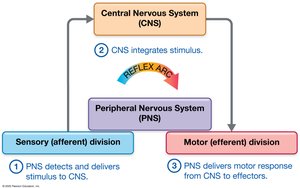
The Peripheral Nervous System (PNS) links the Central Nervous System (CNS) with the rest of the body and the external environment. It is responsible for detecting sensory stimuli and delivering them to the CNS, which processes the input and transmits impulses back through the PNS to effectors (muscle cells and glands) for motor output. Both spinal nerves and cranial nerves are part of the PNS, even though they attach directly to the spinal cord and brain.
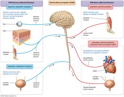
Divisions of the PNS
Sensory (Afferent) Division: Carries sensory information to the CNS.
Somatic Sensory Division: Signals from muscles, bones, joints, skin, and special senses.
Visceral Sensory Division: Signals from internal organs (thoracic and abdominopelvic cavities).
Motor (Efferent) Division: Carries motor commands from the CNS to effectors.
Somatic Motor Division: Signals to skeletal muscles.
Visceral Motor Division (Autonomic Nervous System, ANS): Signals to cardiac and smooth muscles and glands.
Sympathetic Nervous System: "Fight or Flight" responses.
Parasympathetic Nervous System: "Rest and Digest" responses.
Peripheral Nerves and Associated Ganglia
Structure and Function of Peripheral Nerves
Peripheral nerves are bundles of axons from many neurons, bound together by connective tissue sheaths. They innervate most body structures and are classified as:
Mixed nerves: Contain both sensory and motor axons.
Sensory nerves: Contain only sensory axons.
Motor nerves: Mostly motor axons, with some sensory axons monitoring muscle stretch and tension.
Cranial nerves attach to the brain and innervate the head and neck, while spinal nerves branch from the spinal cord and innervate structures below the neck.
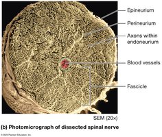
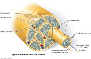
Connective Tissue Sheaths of Spinal Nerves
Epineurium: Surrounds each nerve.
Perineurium: Surrounds each fascicle (group of axons).
Endoneurium: Surrounds each individual axon.
Functional Overview of the PNS
Sensory and Motor Pathways
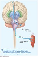
Sensory neurons detect stimuli at sensory receptors and transmit them via spinal or cranial nerves to the CNS.
The CNS processes and integrates the input, then initiates an appropriate motor response via motor areas of the brain.
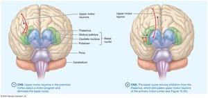
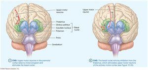
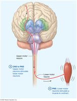
Impulses travel through upper motor neurons to the spinal cord, then through lower motor neurons of the PNS to effectors.
Cranial Nerves
Classification and Functions
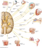
There are twelve pairs of cranial nerves, each with specific sensory, motor, or mixed functions. They are often remembered by mnemonics and classified as:
Sensory Only: Olfactory, Optic, Vestibulocochlear
Motor Only: Oculomotor, Trochlear, Abducens, Accessory, Hypoglossal
Mixed: Trigeminal, Facial, Glossopharyngeal, Vagus
Examples of Cranial Nerve Disorders
Trigeminal Neuralgia: Chronic pain syndrome affecting the trigeminal nerve, causing brief attacks of intense pain. May be triggered by touch or chewing.
Bell’s Palsy: Rapid onset facial nerve impairment, leading to weakness or paralysis of facial muscles, affecting expressions and sometimes taste.

Spinal Nerves and Plexuses
Structure and Branches
Spinal nerves exit the vertebral cavity and split into:
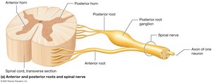
Posterior Ramus: Travels to the posterior side of the body.
Anterior Ramus: Travels to the anterior side and/or limbs.
Ramus Communicans: Contains visceral motor/autonomic axons.
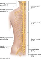
There are 31 pairs of spinal nerves: 8 cervical, 12 thoracic, 5 lumbar, 5 sacral, 1 coccygeal.
Anterior rami of cervical, lumbar, and sacral nerves form plexuses (networks serving muscles and skin).
Cervical Plexus
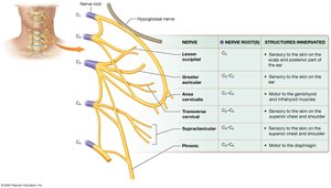
Located deep in the neck, lateral to the first four cervical vertebrae.
Supplies sensory branches to skin of neck, head, chest, and shoulders; motor branches to neck muscles.
Major motor branch: Phrenic nerve (C3, C4, C5) supplies the diaphragm.
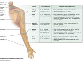
Brachial Plexus
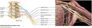
Originates from anterior rami of spinal nerves; innervates the upper limb.
Includes superior, middle, and inferior trunks; splits into anterior and posterior divisions forming cords.
Major nerves: Axillary, Radial, Musculocutaneous, Median, Ulnar.
Lumbar Plexus
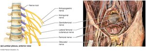
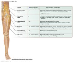
Located anterior to vertebrae, within psoas major muscle; supplies pelvis and lower extremity.
Major nerves: Obturator (anterior division), Femoral (posterior division).
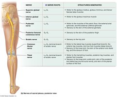
Sacral Plexus
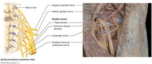
Located inferior to lumbar plexus; supplies pelvis, gluteal region, and lower extremity.
Largest nerve: Sciatic nerve (splits into tibial and common fibular nerves).
Sensory Receptors and Sensation
Types and Mechanisms of Sensory Receptors
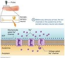
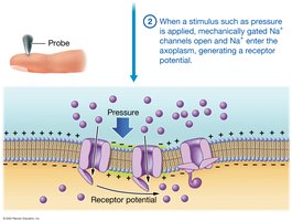
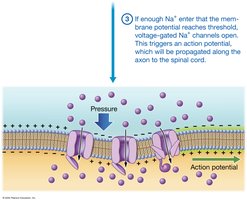
Sensory transduction: Conversion of a stimulus into an electrical signal at a sensory receptor.
Encapsulated nerve endings: Surrounded by supporting cells.
Free nerve endings: Lack supportive cells.
Classification of Sensory Receptors
By Location:
Exteroceptors: Near body surface, detect external stimuli.
Interoceptors: Within body, detect internal stimuli.
By Stimulus Type:
Mechanoreceptors: Respond to mechanical deformation.
Thermoreceptors: Respond to temperature changes.
Chemoreceptors: Respond to chemicals.
Photoreceptors: Respond to light (eye only).
Nociceptors: Detect pain.
Types of Mechanoreceptors
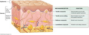
Tactile (Merkel) nerve endings: Discriminative touch, form, and texture.
Tactile (Meissner) corpuscles: Discriminative touch, less fine than Merkel.
Bulbous corpuscles (Ruffini): Stretch and movement.
Lamellated corpuscles (Pacinian): High-frequency vibration and deep pressure.
Hair follicle receptors: Respond to hair bending.
Proprioceptors: Detect movement and position of joints/body parts.
Sensory Neurons and Pathways
Structure and Function of Sensory Neurons
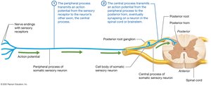
First-order somatic sensory neurons: Pseudounipolar, with cell body in posterior root ganglion, peripheral process (axon), and central process (axon to CNS).
Axons classified by diameter and myelin thickness:
Large diameter, thick myelin: Fast conduction (proprioception, touch).
Small diameter, little myelin: Slow conduction (pain, temperature).
Receptive Fields and Two-Point Discrimination
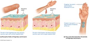
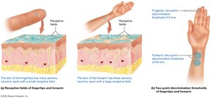
Receptive field: Area served by a neuron.
Large receptive fields: Less sensation, more branching.
Small receptive fields: More sensation, less branching (e.g., fingertips).
Two-point discrimination: Measures relative size of receptive fields.
Dermatomes and Referred Pain
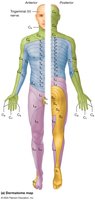
Dermatomes: Skin segments supplied by specific spinal nerves; used clinically to test sensory pathway integrity.
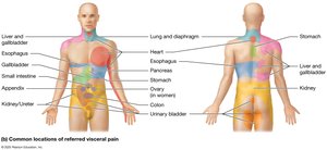
Referred pain: Pain from an organ perceived as cutaneous pain due to shared spinal nerve pathways.
Motor Output and Reflexes
Motor Neurons and Control of Movement
Upper motor neurons: Initiate movement in the primary motor cortex.
Lower motor neurons: Relay messages to muscle fibers, releasing acetylcholine to initiate contraction.
Motor neuron pools: Groups of lower motor neurons innervating the same muscle.
Alpha (α) motor neurons: Stimulate skeletal muscle contraction.
Gamma (γ) motor neurons: Innervate intrafusal fibers (stretch receptors).
Reflex Arcs
Reflex: Programmed, automatic response to sensory input, often protective.
Monosynaptic reflex: Single synapse between sensory and motor neurons.
Polysynaptic reflex: Multiple synapses.
Visceral reflex: Involves internal organs (ANS).
Somatic reflex: Involves skeletal muscles and head structures.
Stretch Receptors in Skeletal Muscles
Muscle spindles: Embedded in contractile muscle fibers; detect stretch and initiate contraction.
Golgi tendon organs: Mechanoreceptors in tendons; monitor tension and prevent damage by causing relaxation.
Types of Somatic Reflexes
Simple stretch reflex: Monosynaptic; restores muscle to optimal length (e.g., patellar reflex).
Flexion (withdrawal) and crossed-extension reflexes: Pull back from painful stimulus and extend opposite limb for balance.
Golgi tendon reflex: Protects muscles/tendons from excessive tension.
Cranial nerve reflexes: Polysynaptic; e.g., gag reflex, corneal blink reflex.
Clinical Disorders of Sensory and Motor Neurons
Peripheral Neuropathies
Sensory neuron disorders: Symptoms depend on affected nerve.
Lower motor neuron disorders: Injury to nerves or cell bodies; may cause paralysis or weakness.
Upper motor neuron disorders: Affect CNS pathways; initial paralysis (spinal shock), followed by spasticity and abnormal reflexes (Babinski sign).
Amyotrophic Lateral Sclerosis (ALS)
Degeneration of α-motor neurons in spinal cord and upper motor neurons in cerebral cortex.
Symptoms: Muscle weakness, upper motor neuron signs, cognitive/behavioral changes.
Death typically within 5 years of onset.
Summary Table: Major Plexuses and Their Nerves
Plexus | Major Nerves | Structures Innervated |
|---|---|---|
Cervical | Phrenic | Diaphragm, skin of neck, head, chest, shoulders |
Brachial | Axillary, Radial, Musculocutaneous, Median, Ulnar | Upper limb muscles and skin |
Lumbar | Obturator, Femoral | Pelvis, anterior and medial thigh, leg, foot |
Sacral | Sciatic (Tibial, Common Fibular) | Hip, pelvis, gluteal region, lower extremity |
Additional info: Academic context was added to clarify the structure and function of the PNS, cranial and spinal nerves, sensory receptors, and clinical disorders. Tables and images were selected strictly for direct relevance to the adjacent content.
