 Back
BackPeripheral Nervous System: Structure, Function, and Clinical Relevance
Study Guide - Smart Notes
 Tailored notes based on your materials, expanded with key definitions, examples, and context.
Peripheral Nervous System (PNS): Overview
Introduction to the PNS
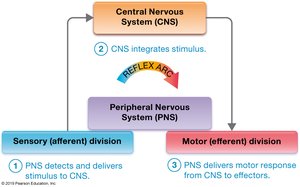
The Peripheral Nervous System (PNS) connects the Central Nervous System (CNS) to the rest of the body and the external environment. It is responsible for detecting sensory stimuli and transmitting information to the CNS, which processes the input and sends motor commands back through the PNS to muscles and glands.
Sensory input: Detection and transmission of stimuli to the CNS.
Motor output: CNS processes input and sends impulses to effectors (muscles/glands).
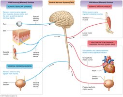
Functional Divisions of the PNS
Sensory (Afferent) Division:
Somatic sensory: Detects internal and external stimuli; general sense receptors (skin), special sense receptors (organs).
Visceral sensory: Relays internal information (e.g., blood pressure) from organs.
Motor (Efferent) Division:
Somatic motor: Voluntary control of skeletal muscles.
Visceral motor (Autonomic Nervous System, ANS): Involuntary control of cardiac muscle, smooth muscle, and glands.
Sympathetic: "Fight or flight" responses.
Parasympathetic: "Rest and digest" functions.
Peripheral Nerves and Associated Ganglia
Structure and Classification of Peripheral Nerves
Peripheral nerves are bundles of axons bound by connective tissue. They innervate most body structures and are classified as:
Mixed nerves: Contain both sensory and motor neurons.
Sensory nerves: Contain only sensory neurons.
Motor nerves: Contain mostly motor neurons (with some sensory neurons for muscle stretch/tension).
Spinal Nerves: Anatomy and Function
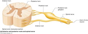
Spinal nerves originate from the spinal cord and innervate structures below the head and neck. They are formed by the fusion of:
Anterior root: Motor neurons from the anterior horn.
Posterior root: Sensory neurons from the posterior horn.
Posterior root ganglion: Houses cell bodies of sensory neurons.
All 31 pairs of spinal nerves are mixed nerves.
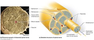
Connective Tissue Layers of Nerves
Epineurium: Outermost layer, holds axons together.
Perineurium: Surrounds fascicles (bundles of axons).
Endoneurium: Surrounds individual axons.
Cranial Nerves
Attach to the brain and innervate head and neck structures.
Can be purely sensory, mixed, or mostly motor.
Cranial Nerves: Classification and Function
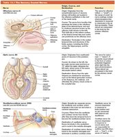
Sensory Cranial Nerves
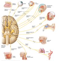
Three cranial nerves are purely sensory:
Olfactory (I): Smell
Optic (II): Vision
Vestibulocochlear (VIII): Hearing and balance
Nerve | Origin | Function |
|---|---|---|
Olfactory (I) | Olfactory epithelium | Smell |
Optic (II) | Retina | Vision |
Vestibulocochlear (VIII) | Inner ear | Hearing, balance |
Motor Cranial Nerves
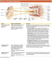
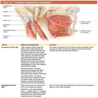
Five cranial nerves are primarily motor:
Oculomotor (III): Eye movement
Trochlear (IV): Eye movement
Abducens (VI): Eye movement
Accessory (XI): Neck muscles
Hypoglossal (XII): Tongue movement
Mixed Cranial Nerves
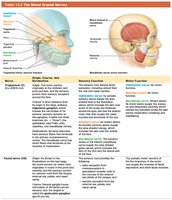
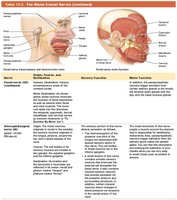
Four cranial nerves contain both sensory and motor axons:
Trigeminal (V): Facial sensation, chewing
Facial (VII): Taste, facial expression
Glossopharyngeal (IX): Taste, swallowing
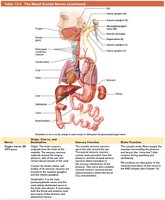
Vagus (X): Visceral sensation, parasympathetic control
Spinal Nerves and Plexuses
Structure and Function of Spinal Nerves
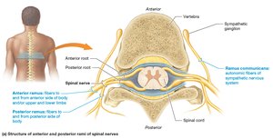
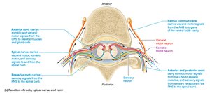
Spinal nerves divide into posterior and anterior rami, carrying somatic motor and sensory information to different regions of the body.
Spinal Nerve Distribution
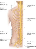
31 pairs: 8 cervical, 12 thoracic, 5 lumbar, 5 sacral, 1 coccygeal
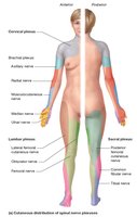
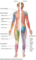
Plexuses: Cervical, brachial, lumbar, sacral
Cervical Plexus
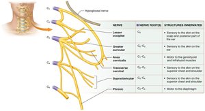
Innervates skin and muscles of neck, head, chest, and shoulders. The phrenic nerve (C3–C5) innervates the diaphragm.
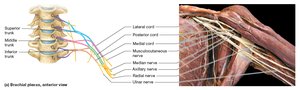
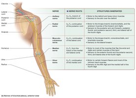
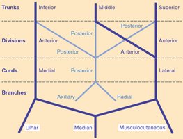
Brachial Plexus
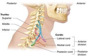
Innervates upper limbs. Major nerves: axillary, radial, musculocutaneous, median, ulnar.
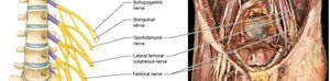
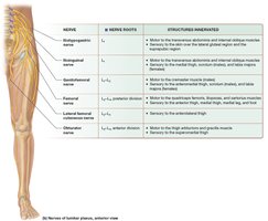
Lumbar Plexus
Innervates pelvic structures and lower extremity. Major nerves: obturator, femoral.
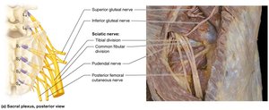
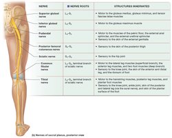
Sacral Plexus
Innervates pelvis, gluteal region, and lower extremity. Major nerves: sciatic, tibial, common fibular.
Cutaneous and Motor Distribution
Sensation: Role of the PNS
Sensory Reception and Transduction
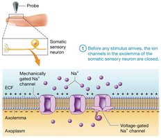
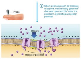
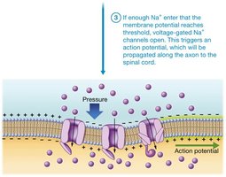
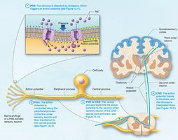
Sensory neurons detect stimuli and transmit them to the CNS. Sensory transduction converts stimuli into electrical signals via ion channels.
Mechanically gated sodium channels open in response to stimulus.
Depolarization (receptor potential) occurs.
If threshold is reached, voltage-gated sodium channels open, generating an action potential.
Adaptation of Sensory Receptors
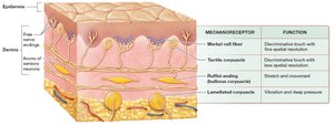
Rapidly adapting: Respond quickly, then stop signaling (e.g., tactile corpuscles).
Slowly adapting: Maintain constant action potentials (e.g., Merkel cell fibers).
Classification of Sensory Receptors
Encapsulated nerve endings: Surrounded by supportive cells.
Free nerve endings: Lack supportive cells.
Exteroceptors: Detect external stimuli.
Interoceptors: Detect internal stimuli.
Mechanoreceptors: Respond to mechanical deformation.
Thermoreceptors: Respond to temperature changes.
Chemoreceptors: Respond to chemical binding.
Photoreceptors: Respond to light (in eye).
Nociceptors: Respond to noxious stimuli (pain).
Somatic Sensory Neurons
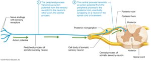
First-order neurons: Pseudounipolar, with cell bodies in dorsal root ganglia.
Peripheral process: Transmits action potentials from receptor to cell body.
Central process: Transmits action potentials to CNS.
Receptive Fields and Two-Point Discrimination
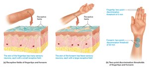
Regions with many small receptive fields (e.g., fingertips) have high sensitivity.
Two-point discrimination threshold measures receptive field size.
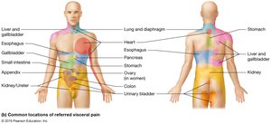
Dermatomes and Referred Pain
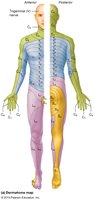
Dermatomes: Skin segments supplied by specific spinal nerves.
Referred pain: Pain from organs perceived as cutaneous pain along dermatome.
Motor Output: Role of the PNS in Movement
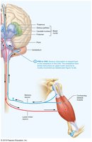
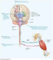
Motor Neurons
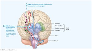
Upper motor neurons: Initiate movement in CNS.
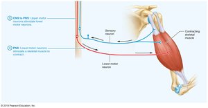
Lower motor neurons: Receive signals and directly stimulate muscle fibers via acetylcholine.
Motor neuron pools: Groups of lower motor neurons innervating the same muscle.
Reflex Arcs: Integration of Sensory and Motor Functions
Reflex Arcs
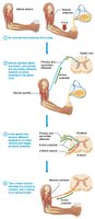
Reflexes are automatic responses to stimuli, typically protective, and involve a three-step sequence: sensory input, CNS integration, and motor output.
Stretch Receptors in Skeletal Muscles
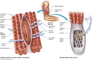
Muscle spindles: Detect muscle stretch and position.
Golgi tendon organs: Monitor tension and protect against excessive force.
Types of Reflexes
Monosynaptic: Single synapse (e.g., patellar reflex).
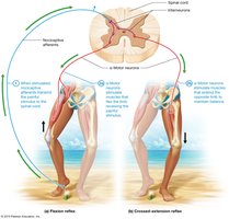
Polysynaptic: Multiple synapses (e.g., withdrawal reflex).
Simple stretch reflex: Muscle contracts in response to stretch.
Golgi tendon reflex: Muscle relaxes in response to excessive tension.
Flexion (withdrawal) reflex: Withdraws limb from painful stimulus.
Crossed-extension reflex: Maintains balance during withdrawal.
Cranial nerve reflexes: Involve cranial nerves (e.g., gag, corneal blink reflex).
Summary Table: Cranial Nerves
Nerve | Type | Main Function |
|---|---|---|
Olfactory (I) | Sensory | Smell |
Optic (II) | Sensory | Vision |
Oculomotor (III) | Motor | Eye movement |
Trochlear (IV) | Motor | Eye movement |
Trigeminal (V) | Mixed | Facial sensation, chewing |
Abducens (VI) | Motor | Eye movement |
Facial (VII) | Mixed | Taste, facial expression |
Vestibulocochlear (VIII) | Sensory | Hearing, balance |
Glossopharyngeal (IX) | Mixed | Taste, swallowing |
Vagus (X) | Mixed | Visceral sensation, parasympathetic control |
Accessory (XI) | Motor | Neck muscles |
Hypoglossal (XII) | Motor | Tongue movement |
Key Equations
Action Potential Generation
Membrane potential change required for action potential:
Ohm's Law in Neural Conduction
Relationship between current, voltage, and resistance:
Conclusion
The Peripheral Nervous System is essential for linking the CNS to the body, enabling sensation, movement, and reflexes. Understanding its structure and function is critical for clinical assessment and treatment of neurological disorders.
