 Back
Peripheral Nervous System: Structure, Function, and Autonomic Regulation
Study Guide - Smart Notes
 Tailored notes based on your materials, expanded with key definitions, examples, and context.
Peripheral Nervous System (PNS)
Overview
The Peripheral Nervous System (PNS) connects the Central Nervous System (CNS) to limbs and organs. It is divided into sensory (afferent) and motor (efferent) divisions. The efferent division is further subdivided into the somatic and autonomic nervous systems, each with distinct structural and functional features.
Reflexes and Homeostasis
Components of a Reflex
Receptor: Detects a stimulus (e.g., pain, temperature).
Sensory neuron: Transmits afferent impulses to the CNS.
Integration center: Processes information in the CNS (spinal cord or brain).
Motor neuron: Conducts efferent impulses from the CNS to an effector.
Effector: Muscle or gland that responds to the motor neuron signal.
Example: The knee-jerk reflex is a classic monosynaptic reflex arc.
Reflexes and Homeostasis
Reflexes help maintain homeostasis by providing rapid, automatic responses to changes in the internal or external environment.
Examples include withdrawal from pain, regulation of heart rate, and control of blood pressure.
Neuromuscular and Neuro-Effector Junctions
Somatic Neuromuscular Junction
Presynaptic cell: Somatic motor neuron
Postsynaptic cell: Skeletal muscle fiber
Neurotransmitter: Acetylcholine (ACh)
Receptor type: Nicotinic cholinergic receptor
Effector cell type: Skeletal muscle
Neurotransmitter removal: Enzymatic degradation by acetylcholinesterase
Parasympathetic Neuro-Effector Junction
Presynaptic cell: Parasympathetic postganglionic neuron
Postsynaptic cell: Target organ (smooth muscle, cardiac muscle, or gland)
Neurotransmitter: Acetylcholine (ACh)
Receptor type: Muscarinic cholinergic receptor
Effector cell type: Smooth muscle, cardiac muscle, or gland
Neurotransmitter removal: Enzymatic degradation by acetylcholinesterase or diffusion
Sympathetic Neuro-Effector Junction
Presynaptic cell: Sympathetic postganglionic neuron
Postsynaptic cell: Target organ (smooth muscle, cardiac muscle, or gland)
Neurotransmitter: Norepinephrine (NE) or epinephrine (Epi)
Receptor type: Adrenergic receptors (alpha or beta)
Effector cell types: Smooth muscle, cardiac muscle, or gland
Neurotransmitter removal: Reuptake into nerve terminal, enzymatic degradation (monoamine oxidase, catechol-O-methyltransferase), or diffusion
Adrenergic and Cholinergic Receptors
Adrenergic Receptors (Sympathetic)
Alpha (α) receptors: Generally cause vasoconstriction and increased peripheral resistance.
Beta (β) receptors: Subdivided into β1 (increases heart rate and contractility), β2 (bronchodilation, vasodilation), and β3 (lipolysis in adipose tissue).
Effect of sympathetic innervation: "Fight or flight" responses—elevated heart rate, dilated pupils, decreased digestive activity, increased blood glucose.
Cholinergic Receptors (Parasympathetic)
Nicotinic receptors: Found at all autonomic ganglia and neuromuscular junctions; always excitatory.
Muscarinic receptors: Found at parasympathetic neuro-effector junctions; can be excitatory or inhibitory depending on the tissue.
Effect of parasympathetic innervation: "Rest and digest" responses—decreased heart rate, increased digestive activity, constricted pupils.
Sympathetic NS and Adrenal Medulla
Anatomical and Functional Relationship
The adrenal medulla is functionally a modified sympathetic ganglion.
Preganglionic sympathetic neurons synapse directly on chromaffin cells in the adrenal medulla, which release epinephrine and norepinephrine into the bloodstream.
This amplifies and prolongs the sympathetic response throughout the body.
Regulation of Autonomic Function
Autonomic function is regulated by the hypothalamus, brainstem, and spinal cord.
Feedback from sensory receptors and higher brain centers modulates autonomic output to maintain homeostasis.
Adrenal Glands: Structure and Development
Structure
Adrenal cortex: Outer region, produces steroid hormones (cortisol, aldosterone, androgens).
Adrenal medulla: Inner region, produces catecholamines (epinephrine and norepinephrine).
Embryological Development
The cortex develops from mesoderm, while the medulla arises from neural crest cells.
Adrenal Medullary Hormones
Epinephrine (adrenaline): Increases heart rate, dilates airways, mobilizes energy stores.
Norepinephrine (noradrenaline): Constricts blood vessels, increases blood pressure.
Stress Responses
Eustress vs. Distress
Eustress: Beneficial, motivating stress (e.g., exercise, achieving goals).
Distress: Harmful, chronic stress (e.g., illness, prolonged anxiety).
Sympathetic/Adrenal (SAM) Response
Acute stress activates the sympathetic nervous system and adrenal medulla, releasing catecholamines for immediate "fight or flight" responses.
Example: Completing a 10K run or undergoing minor surgery triggers increased heart rate, blood pressure, and energy mobilization.
Hypothalamic/Pituitary/Adrenal (HPA) Response
Chronic or severe stress activates the HPA axis, leading to the release of corticotropin-releasing hormone (CRH), adrenocorticotropic hormone (ACTH), and cortisol.
Example: Completing a triathlon or coping with chronic illness results in sustained cortisol release, affecting metabolism and immune function.
Analysis of Peripheral Motor NS Pathways (Diagram)
The diagram below illustrates four branches of the peripheral motor nervous system, each with distinct features:
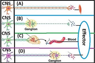
Box | Branch | Signal Molecule | Specific Molecule | Receptor | Synapse | Effector |
|---|---|---|---|---|---|---|
A | Somatic | Neurotransmitter | ACh | Nicotinic | Neuromuscular | Skeletal muscle |
B | Parasympathetic | Neurotransmitter | ACh | Muscarinic | Neuro-effector | Cardiac muscle, smooth muscle, glands |
C | Sympathetic (Adrenal Medulla/SAM) | Hormone | Epinephrine, Norepinephrine | Alpha, Beta | Neuro-effector (via blood) | Cardiac muscle, smooth muscle, glands |
D | Sympathetic | Neurotransmitter | Norepinephrine | Alpha, Beta | Neuro-effector | Cardiac muscle, smooth muscle, glands |
Additional info: The table summarizes the key features of each branch, including the type of signal molecule, receptor, synapse, and effector involved.
