 Back
BackPeripheral Nervous System: Structure, Function, and Cranial Nerves
Study Guide - Smart Notes
 Tailored notes based on your materials, expanded with key definitions, examples, and context.
The Peripheral Nervous System (PNS)
Overview and Components
The Peripheral Nervous System (PNS) connects the central nervous system (CNS) to limbs and organs, serving as a communication relay. It is divided into sensory and motor divisions, each with distinct functions.
Sensory Division: Transmits impulses from receptors in the skin, muscles, joints (somatic afferents), and visceral organs (visceral afferents) to the CNS.
Motor Division: Efferent fibers carry impulses from the CNS to effectors such as muscles, glands, and viscera.
Spinal Nerves: 31 pairs connect the spinal cord to the body.
Cranial Nerves: 12 pairs connect the brain to the head, neck, and some internal organs.

Motor Division of the PNS
The motor division is further subdivided into the somatic and autonomic nervous systems.
Somatic Nervous System: Controls voluntary movements by conducting impulses to skeletal muscles.
Autonomic Nervous System: Regulates involuntary activities in smooth muscle, cardiac muscle, and glands. It has two subdivisions:
Sympathetic N.S.: Responsible for 'fight or flight' responses.
Parasympathetic N.S.: Responsible for 'resting and digesting' activities.
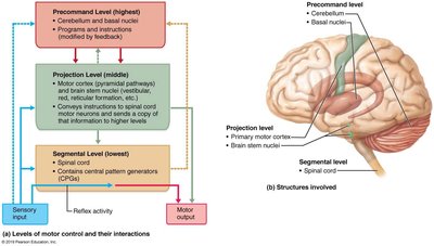
Levels of Motor Control
Motor activity is regulated at three hierarchical levels: segmental, projection, and precommand.
Segmental Level: Spinal reflexes and central pattern generators (CPGs) in the spinal cord.
Projection Level: Motor cortex and brainstem nuclei, which send instructions to the spinal cord.
Precommand Level: Cerebellum and basal nuclei, which plan and coordinate movements.
Sensory Division of the PNS
Sensory Neural Integration
Sensory information is processed at three levels:
Receptor Level: Sensory receptors generate action potentials in response to stimuli.
Circuit Level: Ascending pathways relay signals through first, second, and third order neurons.
Perceptual Level: Cerebral cortex integrates sensory inputs for perception.
Sensory Receptors
Sensory receptors are classified by location and the type of stimuli they detect.
Location:
Exteroceptors: Near the body surface; detect external stimuli (touch, pressure, pain, temperature, special senses).
Interoceptors (Visceroceptors): Detect internal stimuli (pain, discomfort, tissue stretch, temperature).
Proprioceptors: Monitor internal stimuli in muscles, tendons, ligaments, and joints; sense movement and stretch.
Stimulus Type:
Mechanoreceptors: Respond to mechanical forces (touch, pressure, vibration).
Chemoreceptors: Detect dissolved chemicals (smell, taste).
Photoreceptors: Detect light energy (vision).
Thermoreceptors: Detect temperature changes.
Nociceptors: Detect pain or potentially damaging stimuli.
General Sensory Receptors: Structure and Function
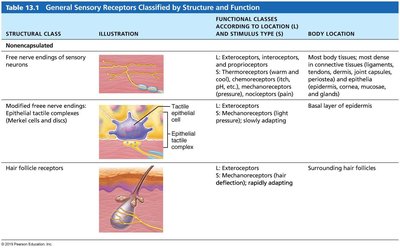
Sensory receptors are further classified as encapsulated or nonencapsulated, each with specific functions and locations.
Structural Class | Illustration | Functional Class | Body Location |
|---|---|---|---|
Free nerve endings of sensory neurons | — | Exteroceptors, interoceptors, proprioceptors; thermoreceptors, chemoreceptors, mechanoreceptors, nociceptors | Most body tissues, especially connective tissues and epithelia |
Modified free nerve endings (Merkel discs) | — | Exteroceptors; mechanoreceptors (pressure), slowly adapting | Basal layer of epidermis |
Hair follicle receptors | — | Exteroceptors; mechanoreceptors (hair deflection), rapidly adapting | Surrounding hair follicles |
Structural Class | Illustration | Functional Class | Body Location |
|---|---|---|---|
Tactile (Meissner's) corpuscles | — | Exteroceptors; mechanoreceptors (light pressure), rapidly adapting | Dermal papillae of hairless skin |
Lamellar (Pacinian) corpuscles | — | Exteroceptors, interoceptors, some proprioceptors; mechanoreceptors (deep pressure, vibration), rapidly adapting | Dermis, hypodermis, periosteum, mesentery, etc. |
Bulbous (Ruffini) endings | — | Exteroceptors, proprioceptors; mechanoreceptors (deep pressure, stretch), slowly adapting | Dermis, hypodermis, joint capsules |
Muscle spindles | — | Proprioceptors; mechanoreceptors (muscle stretch) | Skeletal muscles |
Tendon organs | — | Proprioceptors; mechanoreceptors (tendon stretch) | Tendons |
Joint kinesthetic receptors | — | Proprioceptors; mechanoreceptors, nociceptors | Joint capsules of synovial joints |

Cranial Nerves
Overview of Cranial Nerves
Cranial nerves are twelve pairs of nerves that emerge directly from the brain. They serve the head, neck, and some internal organs, and may contain sensory, motor, or parasympathetic fibers.
C.N. (I) and (II): Originate from the cerebrum.
C.N. (III)–(XII): Originate from the brainstem.
C.N. (X) Vagus: Extends into the abdominal cavity.
Parasympathetic fibers: Present in C.N. (III), (VII), (IX), and (X).

Cranial Nerve | Sensory Function | Motor Function | Parasympathetic Fibers |
|---|---|---|---|
I Olfactory | Yes | No | No |
II Optic | Yes | No | No |
III Oculomotor | No | Yes | Yes |
IV Trochlear | No | Yes | No |
V Trigeminal | Yes | Yes | No |
VI Abducens | No | Yes | No |
VII Facial | Yes | Yes | Yes |
VIII Vestibulocochlear | Yes | No | No |
IX Glossopharyngeal | Yes | Yes | Yes |
X Vagus | Yes | Yes | Yes |
XI Accessory | No | Yes | No |
XII Hypoglossal | No | Yes | No |
Selected Cranial Nerves
C.N. I: Olfactory Nerves
The olfactory nerves are purely sensory and carry sensations of smell from the nasal cavity to the brain.
Originate in the mucosa of the nasal cavity.
Pass through the cribiform plate of the ethmoid bone to the olfactory bulb.
Pathway: Olfactory nerves → olfactory bulb → olfactory tract → inferior frontal and medial temporal lobes.


C.N. II: Optic Nerves
The optic nerves are sensory only and originate from the retina.
Pathway: Optic nerves → optic chiasma → optic tracts → lateral geniculate bodies of thalamus → optic radiations → visual cortex in occipital lobe.

C.N. III: Oculomotor Nerves
The oculomotor nerves contain motor and parasympathetic fibers.
Originate from the infero-ventral midbrain.
Motor to extrinsic eye muscles: inferior oblique, superior rectus, inferior rectus, medial rectus.
Parasympathetic fibers to constrictor pupillary muscles and ciliary muscles (constrict pupils, thicken lens).

C.N. IV: Trochlear Nerves
Motor only.
From infero-dorsal midbrain; motor to the superior oblique muscle.
C.N. V: Trigeminal Nerves
Motor and sensory; largest cranial nerve.
Three branches:
Ophthalmic (V1): Sensory from upper eyelid, eye surface, tear glands, nose, scalp, forehead.
Maxillary (V2): Sensory from upper teeth, gum, lip, palate, cheek, lower eyelid.
Mandibular (V3): Motor to muscles of mastication; sensory from lower teeth, gum, lip, jaw, scalp.
C.N. VI: Abducens Nerves
Motor only; leave inferior pons; motor to lateral rectus muscles.
C.N. VII: Facial Nerves
Motor, sensory, and parasympathetic fibers.
Motor to muscles of facial expression.
Taste from anterior 2/3 of tongue.
Parasympathetic innervation: salivary glands (submandibular, sublingual), lacrimal glands.
C.N. VIII: Vestibulocochlear Nerves
Sensory only; pons-medulla border.
Cochlear branch: auditory messages.
Vestibular branch: equilibrium information.
C.N. IX: Glossopharyngeal Nerves
Motor, sensory, and parasympathetic fibers.
Motor to pharynx for swallowing.
Taste from posterior 1/3 of tongue; sensory from tongue, tonsils, eustachian tubes, carotid arteries.
Parasympathetic to parotid salivary glands.
C.N. X: Vagus Nerves
Motor, sensory, and parasympathetic fibers.
Motor to pharynx and larynx for swallowing.
Sensory from posterior tongue, pharynx, thoracic and abdominal viscera.
Parasympathetic to heart, lungs, thoracic and abdominal viscera.
Only cranial nerve to pass beyond head and neck.
C.N. XI: Spinal Accessory Nerves
Motor only; lateral medulla oblongata.
Motor to shoulder, neck, pharynx, larynx muscles (sternocleidomastoid, trapezius).
C.N. XII: Hypoglossal Nerves
Motor only; medulla oblongata.
Motor to intrinsic and extrinsic tongue muscles.
Summary Table: Cranial Nerves and Functions
Nerve | Function |
|---|---|
Olfactory (I) | Sensory: Smell |
Optic (II) | Sensory: Vision |
Oculomotor (III) | Motor: Eye movement; Parasympathetic: Pupil constriction, lens shape |
Trochlear (IV) | Motor: Eye movement (superior oblique) |
Trigeminal (V) | Sensory: Face; Motor: Mastication |
Abducens (VI) | Motor: Eye movement (lateral rectus) |
Facial (VII) | Motor: Facial expression; Sensory: Taste; Parasympathetic: Salivary, lacrimal glands |
Vestibulocochlear (VIII) | Sensory: Hearing, equilibrium |
Glossopharyngeal (IX) | Motor: Pharynx; Sensory: Taste, blood chemistry; Parasympathetic: Parotid gland |
Vagus (X) | Motor: Pharynx, larynx; Sensory: Viscera; Parasympathetic: Heart, lungs, viscera |
Accessory (XI) | Motor: Neck, shoulder |
Hypoglossal (XII) | Motor: Tongue |
Key Terms and Concepts
Afferent fibers: Carry sensory information to the CNS.
Efferent fibers: Carry motor commands from the CNS to effectors.
Parasympathetic fibers: Regulate involuntary functions such as digestion and heart rate.
Mechanoreceptors: Detect mechanical changes.
Proprioceptors: Sense body position and movement.
Equations and Pathways
Action Potential Generation: (Ohm's Law for neural conduction)
Ascending Pathways:
Additional info:
Tables and diagrams were recreated and expanded for clarity and completeness.
Some cranial nerve details were inferred from standard textbook knowledge.
