Back
BackPhysiology of Smell and Taste: Structure, Function, and Neural Pathways
Study Guide - Smart Notes
 Tailored notes based on your materials, expanded with key definitions, examples, and context.
Physiology of Smell (Olfaction)
Chemical Senses: Overview
The chemical senses—gustation (taste) and olfaction (smell)—are specialized for detecting chemicals dissolved in aqueous solutions. Olfactory chemoreceptors respond to substances dissolved in the fluids of the nasal membranes, while gustatory chemoreceptors respond to substances dissolved in saliva.
Anatomy and Function of the Olfactory System
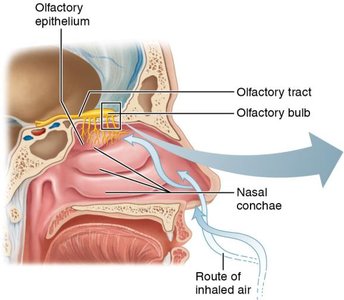
The olfactory system is responsible for detecting and processing odorants. The olfactory epithelium, located in the dorsum of the nasal cavity, contains olfactory receptor neurons, supporting cells, and basal cells. Airflow over the nasal conchae ensures even distribution of odorants across the olfactory epithelium.
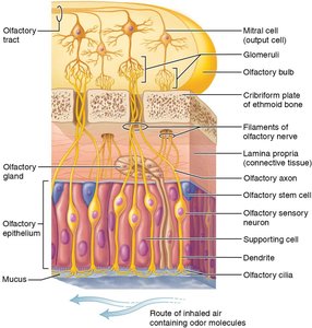
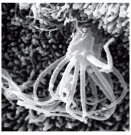
Olfactory receptor neurons are true neurons with dendrites ending in cilia that detect odorants.
Supporting cells maintain the structure and function of the epithelium.
Basal cells are progenitor cells that regenerate olfactory neurons every 4–8 weeks.
Olfactory Receptors and Signal Transduction
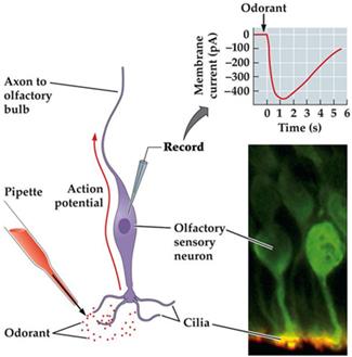
Olfactory cilia contain numerous receptor types, allowing humans to distinguish approximately 10,000 different odors. Odorant molecules bind to specific receptors on the cilia, initiating a cascade that leads to neuronal depolarization and action potential generation.
Each olfactory receptor neuron expresses only one type of receptor, ensuring specificity.
Signal amplification occurs via G protein-coupled receptors, leading to the production of many second messengers (cAMP).
Depolarization is caused by the influx of Na+ and Ca2+ ions through cAMP-gated channels.
Olfactory Signal Transmission and Pathways
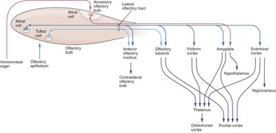
Olfactory receptor neuron axons pass through the cribriform plate to the olfactory bulb, where they synapse with mitral and tufted cells in structures called glomeruli. Each glomerulus receives input from neurons expressing the same receptor type, ensuring specificity in odor detection.
Mitral and tufted cells relay signals via the olfactory tract to the primary olfactory cortex (piriform cortex) and limbic structures.
Unlike other sensory systems, olfactory signals initially bypass the thalamus, projecting directly to cortical and limbic areas.
Further integration occurs in the thalamus and other brain regions, linking smell to memory and emotion.

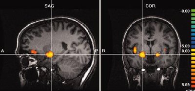
Functional MRI of Olfactory Activation
Functional MRI studies show that olfactory stimulation activates the amygdala, insular cortex, and orbitofrontal cortex. These regions are involved in emotional processing, integration of sensory information, and conscious odor perception.
Bilateral amygdala activation reflects the emotional impact of odors.
Insular and orbitofrontal cortex activation is associated with complex processing and discrimination of odors.
Physiology of Taste (Gustation)
Overview of Taste Perception
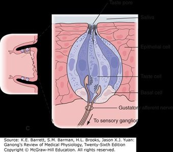
Taste perception requires that substances be dissolved in saliva, allowing them to interact with gustatory hairs on taste cells. The binding of tastants to these hairs depolarizes the taste cell, leading to neurotransmitter release and activation of sensory neurons.
Taste cells transduce chemical signals, while sensory neurons transmit these signals to the brainstem.
Saliva and serous gland secretions facilitate tastant dissolution and receptor interaction.
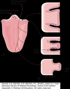
Taste Bud Distribution and Structure
Taste buds are sensory organs for taste, located primarily in papillae on the tongue. There are three main types of papillae: fungiform, circumvallate, and foliate. Each taste bud contains gustatory epithelial cells (sensory), supporting cells, and basal cells (stem cells).
Fungiform papillae: Numerous, at the tip and sides of the tongue.
Circumvallate papillae: Arranged in a V-shape at the back of the tongue.
Foliate papillae: On the lateral edges, important for sour taste detection.
Taste buds are replaced approximately every 7 days by basal cell division.
The Five Basic Taste Sensations
There are five primary taste modalities, each with distinct receptor mechanisms:
Sweet: Triggered by sugars, artificial sweeteners, and some amino acids.
Salt: Initiated by metal ions (e.g., Na+).
Sour: Caused by hydrogen ions (acidity).
Bitter: Activated by alkaloids (e.g., quinine, nicotine).
Umami: Elicited by glutamate, common in protein-rich foods.
Each taste type plays a role in food selection and nutritional regulation. For example, umami detection is linked to protein intake, while bitter detection may help avoid toxins.
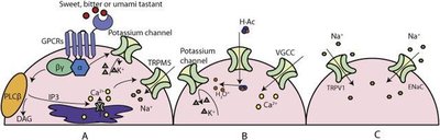
Mechanisms of Taste Receptor Activation
Different taste modalities are transduced by specific cellular mechanisms:
Salty: Na+ enters through ENaC or TRPV1 channels, causing depolarization.
Sour: H+ ions block potassium channels and increase intracellular Ca2+, leading to neurotransmitter release.
Sweet, Bitter, Umami: Bind to G protein-coupled receptors (GPCRs), activating second messenger pathways that result in cell depolarization and neurotransmitter release.
Neural Pathways for Taste
Taste information is transmitted to the brain via three cranial nerves:
Facial nerve (VII): Anterior two-thirds of the tongue.
Glossopharyngeal nerve (IX): Posterior one-third of the tongue.
Vagus nerve (X): Epiglottis and lower pharynx.
All taste signals are relayed to the solitary nucleus in the medulla, then to the thalamus, and finally to the gustatory cortex in the insula. Integration with the limbic system allows for emotional and autonomic responses to taste.
Interconnection Between Taste and Smell
Taste perception is heavily reliant on olfaction; about 80% of what is perceived as taste is attributed to smell. Aerosols produced in the mouth carry flavor compounds to the olfactory epithelium, significantly contributing to flavor perception. Additional sensory inputs such as thermoreceptors and mechanoreceptors on the tongue also influence the overall taste experience.
Genetic Variation in Taste Perception: The Panda Example
Genetic mutations can affect taste perception. For example, pandas have mutations in both copies of the T1R1 gene, which is responsible for umami detection. This may explain their strict bamboo diet despite being potential carnivores.