 Back
BackPhysiology of the Nervous System: Membrane Potentials, Action Potentials, and Synaptic Transmission
Study Guide - Smart Notes
 Tailored notes based on your materials, expanded with key definitions, examples, and context.
Physiology of the Nervous System: Membrane Potentials, Action Potentials, and Synaptic Transmission
Introduction
The nervous system relies on the unique properties of neurons to generate, conduct, and transmit electrical signals. These processes are fundamental for communication within the body, enabling sensation, movement, and cognition. This guide reviews the mechanisms underlying membrane potentials, action potentials, and synaptic transmission, as well as the roles of neurotransmitters and neural circuits.
Basic Principles of Electricity in Neurons
Electrical Properties of Cells
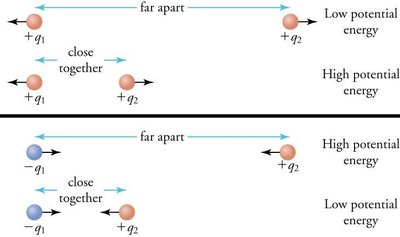
Opposite charges attract, and energy is required to keep them separated across a membrane.
When separated, the system has potential energy (voltage).
Movement of charges (ions) across the membrane generates current.
Key Definitions
Voltage (V): The measure of potential energy generated by separated charge, measured in volts (V) or millivolts (mV).
Current (I): The flow of electrical charge (ions) between two points; can be used to do work.
Resistance (R): Hindrance to charge flow; insulators have high resistance, conductors have low resistance.
Membrane Potential and Ion Gradients
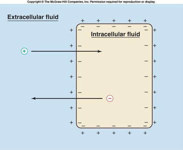
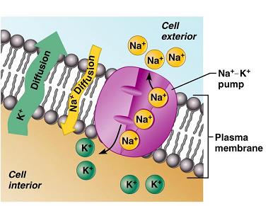
Neurons maintain a resting membrane potential (typically -70 mV) due to differences in ion concentrations inside and outside the cell.
The sodium-potassium ATPase pump (Na+/K+ pump) actively transports 3 Na+ out and 2 K+ in, maintaining the gradient.
Membrane Ion Channels
Types of Ion Channels
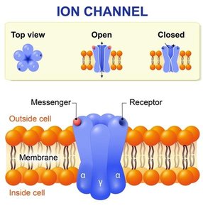
Leakage (nongated) channels: Always open, allowing ions to move along their gradients.
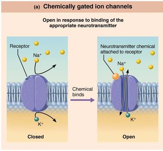
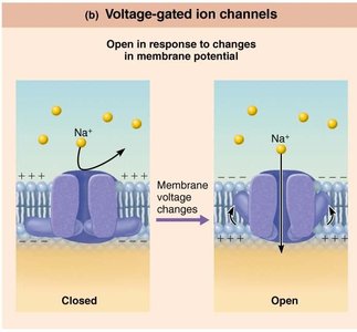
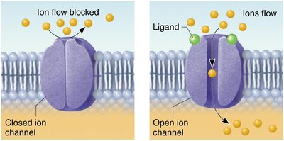
Gated channels: Open or close in response to specific signals.
Chemically gated (ligand-gated): Open with binding of a specific chemical (e.g., neurotransmitter).
Voltage-gated: Open/close in response to changes in membrane potential.
Mechanically gated: Open/close in response to physical deformation (e.g., touch receptors).
Resting Membrane Potential
Establishment of Resting Potential
Resting potential is determined by the concentration gradients of Na+, K+, and Cl-, and the membrane's permeability to these ions.
Potassium (K+) has the greatest influence due to high membrane permeability.
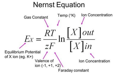
The Nernst equation calculates the equilibrium potential for a single ion:
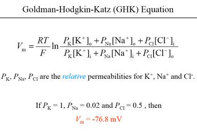
The Goldman-Hodgkin-Katz equation calculates the membrane potential considering multiple ions:
Changes in Membrane Potential
Depolarization and Hyperpolarization
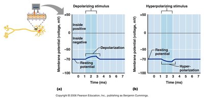
Depolarization: Membrane potential becomes less negative (moves toward zero or positive).
Hyperpolarization: Membrane potential becomes more negative (moves further from zero).
These changes are the basis for neural signaling.
Graded Potentials
Characteristics of Graded Potentials
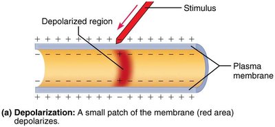
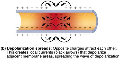
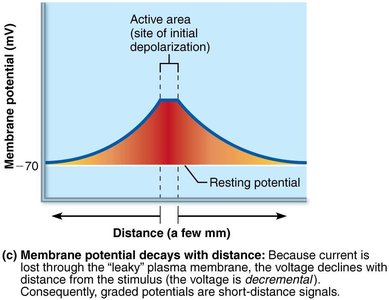
Short-lived, localized changes in membrane potential.
Triggered by a stimulus that opens gated ion channels.
Can be depolarizing or hyperpolarizing.
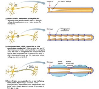
Decay with distance due to current leakage across the membrane.
Types include receptor potentials (in sensory neurons) and postsynaptic potentials (at synapses).
Action Potentials
Mechanism of Action Potentials
Principal means of long-distance neural communication.
Brief reversal of membrane potential (~100 mV change).
Generated by opening of voltage-gated Na+ and K+ channels.
Self-propagating and do not decay with distance.
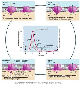
Phases of the Action Potential
Resting state: All gated Na+ and K+ channels closed.
Depolarization: Na+ channels open, Na+ influx.
Repolarization: Na+ channels inactivate, K+ channels open, K+ efflux.
Hyperpolarization: Some K+ channels remain open, Na+ channels reset.
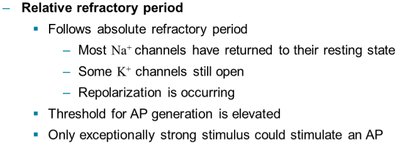
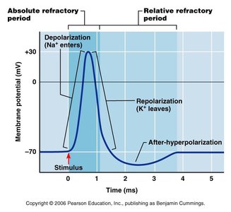
Refractory Periods
Absolute refractory period: No new action potential can be generated, regardless of stimulus strength.
Relative refractory period: A stronger-than-usual stimulus can initiate another action potential.
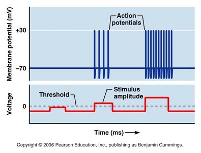
Coding for Stimulus Intensity
All action potentials are identical in amplitude.
Stimulus intensity is encoded by the frequency of action potentials (number per second).
Conduction Velocity
Factors Affecting Conduction Speed
Axon diameter: Larger diameter = faster conduction (less resistance).
Degree of myelination: Myelinated axons conduct impulses faster via saltatory conduction (jumps between nodes of Ranvier).
Classification of Nerve Fibers
Group A: Largest diameter, myelinated, fastest (150 m/s); somatic sensory and motor fibers.
Group B: Intermediate diameter, lightly myelinated (15 m/s); autonomic fibers.
Group C: Smallest diameter, unmyelinated (1 m/s); autonomic fibers.
Synaptic Transmission
The Synapse
Junction where information is transferred from one neuron to another, or to an effector cell (muscle/gland).
Most synapses are chemical synapses using neurotransmitters.
Excitatory and Inhibitory Synapses
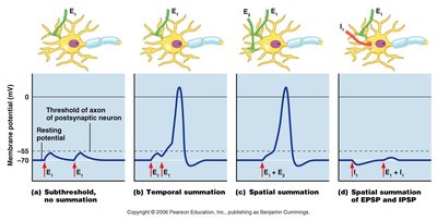
Excitatory postsynaptic potential (EPSP): Neurotransmitter binding causes depolarization, increasing likelihood of action potential.
Inhibitory postsynaptic potential (IPSP): Neurotransmitter binding causes hyperpolarization, decreasing likelihood of action potential.
Neurotransmitter Actions and Receptors
Direct action: Neurotransmitter binds directly to and opens ion channels (e.g., ACh, amino acids).
Indirect action: Neurotransmitter acts through second messengers (e.g., G protein-coupled receptors), causing longer-lasting effects.
Major Neurotransmitters
GABA: Inhibitory, CNS (cortex, retina).
Glutamate: Excitatory, CNS (cortex).
Acetylcholine (ACh): Excitatory or inhibitory, CNS and PNS.
Norepinephrine: Excitatory or inhibitory, CNS and sympathetic nervous system.
Dopamine: CNS, excitatory or inhibitory depending on receptor.
Serotonin: Inhibitory, CNS (sleep).
Histamine: CNS (wakefulness).
Substance P: Pain neurotransmitter.
Enkephalin (endorphin): Pain-blocking neurotransmitter.
Nitric oxide (NO): Excitatory, CNS and PNS, involved in memory.
Neuronal Pools and Neural Circuits
Neuronal Pools
Functional groups of neurons that integrate and forward information.
Patterns of Neural Processing
Serial processing: Input travels along one pathway to a specific destination (e.g., reflex arc).
Parallel processing: Input travels along several pathways, allowing for complex responses and higher-level processing.
Types of Neural Circuits
Diverging circuit: One input, many outputs (amplifies signal).
Converging circuit: Many inputs, one output (concentrates signal).
Reverberating circuit: Signal travels through a chain of neurons, each feeding back to previous neurons (oscillations, rhythmic activity).
Parallel after-discharge circuit: One input, diverges to several pathways, then converges to a single output (complex processing).
Additional info: This guide covers the core physiological principles of neural signaling, including the ionic basis of membrane potentials, the generation and propagation of action potentials, synaptic transmission, and the integration of signals in neural circuits. Understanding these mechanisms is essential for further study of the nervous system and its role in human physiology.
