Back
BackPhysiology of the Nervous System: Motor System and Motor Control
Study Guide - Smart Notes
 Tailored notes based on your materials, expanded with key definitions, examples, and context.
Motor System Overview
Introduction to Motor Systems
The motor system is responsible for the initiation, coordination, and execution of voluntary and involuntary movements. It integrates sensory input, processes motor commands, and regulates muscle activity through a hierarchical network of neural structures.
Levels of Motor Control: Motor control is organized into hierarchical levels: precommand (planning and modulation), projection (relay and execution), and segmental (spinal reflexes and pattern generation).
Key Structures: The cerebral cortex, basal ganglia, cerebellum, brainstem, and spinal cord all contribute to motor function.
Cerebral Cortex: Key Areas in Motor Control and Planning
Functional Regions and Their Roles
The cerebral cortex contains specialized regions that contribute to the planning, initiation, and execution of voluntary movements.
Primary Motor Cortex: Located in the precentral gyrus, it executes voluntary movements and receives sensory feedback for fine-tuning.
Premotor Cortex: Involved in planning and coordinating complex movements, influencing both direct and indirect motor pathways.
Supplementary Motor Area: Coordinates bilateral movements and is active during mental rehearsal of movement sequences.
Frontal Eye Field: Controls voluntary eye movements.
Broca's Area: Specializes in speech production and motor planning for language.
Basal Ganglia: Modulates movement initiation and amplitude.
Cerebellum: Refines movement coordination, timing, and precision.

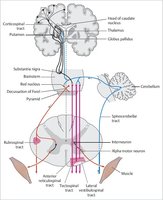
Descending Motor Pathways
Direct and Indirect Pathways
Descending motor pathways transmit commands from the brain to the spinal cord, controlling voluntary and involuntary movements.
Direct (Pyramidal) Pathways: Originate from the motor cortex and project directly to the spinal cord, controlling skilled voluntary movements.
Indirect (Extrapyramidal) Pathways: Involve brainstem nuclei and modulate posture, muscle tone, and automatic movements.
Decussation: Most motor fibers cross at the medullary pyramids, resulting in contralateral control.
Upper Motor Neurons: Originate in the cortex; damage leads to spastic paralysis.
Lower Motor Neurons: Located in the spinal cord or cranial nerve nuclei; damage leads to flaccid paralysis.

The Direct (Pyramidal) Motor Pathway
Structure and Function
The direct pathway, also known as the pyramidal tract, is essential for voluntary, skilled movements.
Origin: Layer V pyramidal neurons in the precentral gyrus.
Pathway: Fibers travel via the corticospinal tract, decussate in the medulla, and synapse on anterior horn cells in the spinal cord.
Corticobulbar Tract: Controls head and neck muscles via cranial nerves.
Lateral Corticospinal Tract: 90% of fibers, cross in the brainstem, control limbs.
Anterior Corticospinal Tract: 10% of fibers, bilateral innervation of midline muscles.

Motor Units and Neuromuscular Innervation
Motor Unit Structure and Function
A motor unit consists of a single motor neuron and all the muscle fibers it innervates. The neuromuscular junction is the site where the motor neuron communicates with the muscle fiber.
Acetylcholine Release: Triggers depolarization and muscle contraction.
Fine vs. Gross Control: Fewer fibers per unit for fine control (e.g., eye muscles); more for gross control (e.g., quadriceps).
One-to-One Innervation: Each muscle fiber is innervated by only one motor neuron.

Types of Paralysis from Spinal Cord Trauma
Clinical Manifestations
Spinal cord injuries can result in different types of paralysis depending on the location and extent of the damage.
Paraplegia: Loss of motor and sensory function in lower limbs (T1-L1 injury).
Quadriplegia: Loss of function in all four limbs (cervical injury).
Spastic Paralysis: Upper motor neuron damage; increased muscle tone and reflexes.
Flaccid Paralysis: Lower motor neuron damage; loss of muscle tone and reflexes.

Indirect (Extrapyramidal) Motor System
Components and Functions
The indirect motor system includes all descending motor pathways outside the pyramidal tracts, modulating posture, balance, and automatic movements.
Rubrospinal Tract: Controls distal limb muscles.
Vestibulospinal Tract: Maintains head position and balance.
Reticulospinal Tract: Regulates posture and reflexes.
Tectospinal Tract: Mediates head and eye movements in response to stimuli.
Integration: Works with the cortex for voluntary actions and coordinates with direct pathways.
Basal Ganglia's Role in Movement Initiation
Function and Clinical Relevance
The basal ganglia are essential for initiating voluntary movement and regulating movement amplitude and direction.
Components: Caudate nucleus, putamen, globus pallidus, substantia nigra, subthalamic nucleus.
Dopamine: Produced by substantia nigra; critical for normal function.
Parkinson's Disease: Caused by degeneration of dopamine-producing neurons, leading to tremors, rigidity, and bradykinesia.

Cerebellar Function in Motor Coordination
Coordination and Feedback
The cerebellum refines and coordinates voluntary movements by integrating sensory input and motor commands.
Inputs: Receives signals from the cortex and sensory systems.
Outputs: Sends corrective feedback to the motor cortex via the thalamus.
Damage: Leads to ataxia and dysmetria (impaired coordination).
Distinction: Basal ganglia initiate movement; cerebellum coordinates and refines it.

Cortico-Cerebellar Communication Pathways
Information Flow and Feedback
Cortico-cerebellar pathways are essential for the integration and correction of motor commands, ensuring precise movement execution.
Pontine Nuclei: Relay sensory and motor information from the cortex to the cerebellum.
Thalamic Relay: Corrective feedback from the cerebellum is sent to the cortex via the ventral lateral nucleus of the thalamus.
Double-Crossed Pathways: Cerebellar output affects the ipsilateral body, refining ongoing movements.

Hierarchical Organization of Motor Control
Levels of Motor Control
Motor control is organized into three hierarchical levels, each with distinct roles in movement planning and execution.
Precommand Level: Cerebellum and basal nuclei plan and modulate movements.
Projection Level: Primary motor cortex and brainstem nuclei relay instructions to the spinal cord.
Segmental Level: Spinal cord mediates reflexes and rhythmic movements via central pattern generators.

The Final Common Pathway of Motor Control
Integration and Execution
Lower motor neurons serve as the final common pathway, integrating all motor signals and executing muscle contractions.
Integration: Inputs from upper motor neurons, cerebellum, basal nuclei, and thalamus converge on lower motor neurons.
Execution: Lower motor neurons transmit the final command to skeletal muscles.

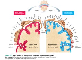
Cortical Representation of Motor and Sensory Functions
Somatotopic Organization
The primary motor and somatosensory cortices are organized somatotopically, with different body regions represented proportionally to their functional importance.
Motor Homunculus: Larger cortical areas are devoted to regions requiring fine motor control (e.g., hands, face).
Sensory Homunculus: Similar proportional representation for sensory input.