 Back
BackProtection and Organization of the Nervous System: Meninges, CSF, Blood-Brain Barrier, and Spinal Cord
Study Guide - Smart Notes
 Tailored notes based on your materials, expanded with key definitions, examples, and context.
Protection of the Central Nervous System (CNS)
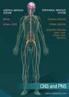
Overview of CNS and PNS
The nervous system is divided into the Central Nervous System (CNS), consisting of the brain and spinal cord, and the Peripheral Nervous System (PNS), which includes cranial nerves, spinal nerves, and sensory organs. The CNS is protected by several anatomical and physiological barriers that ensure its proper function and defense against injury and infection.
Protection of the Brain
Bones, Meninges, Cerebrospinal Fluid, and Blood-Brain Barrier
The brain is protected by four main structures: the cranial bones, the meninges, cerebrospinal fluid (CSF), and the blood-brain barrier. Each plays a unique role in safeguarding neural tissue from mechanical injury, infection, and chemical fluctuations.
Meninges
Structure and Function
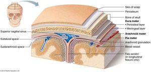
The meninges are three connective tissue membranes that cover and protect the CNS, enclose blood vessels, contain CSF, and form partitions within the skull. The three layers are:
Dura mater: The toughest, outermost layer, consisting of periosteal and meningeal layers. These layers are fused except where they separate to form dural sinuses.
Arachnoid mater: The middle, web-like layer, separated from the dura by the subdural space. The subarachnoid space beneath it contains CSF and major blood vessels.
Pia mater: The innermost, delicate layer that clings tightly to the brain and follows its contours, containing many tiny blood vessels.
Dural Septa and Cranial Partitions
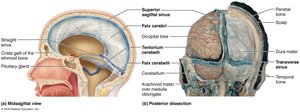
The dura mater forms inward extensions called dural septa that partition the cranial cavity and limit excessive movement of the brain. Major septa include the falx cerebri, falx cerebelli, and tentorium cerebelli.
Clinical Correlates
Meningitis: Inflammation of the meninges, usually due to infection.
Encephalitis: Inflammation of the brain, often viral or autoimmune in origin.
Recent research suggests a possible fourth meningeal membrane, the subarachnoid lymphatic-like membrane (SLYM), which may further compartmentalize the subarachnoid space.
Cerebrospinal Fluid (CSF)
Functions and Composition
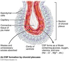
CSF is a clear, colorless liquid that cushions the CNS, provides buoyancy, removes waste, and delivers nutrients. It is similar to plasma but contains less protein and different concentrations of ions (more vitamin C, Na+, Cl-, Mg2+, H+; less Ca2+, K+).
Production and Circulation
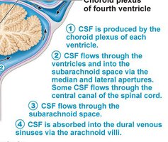
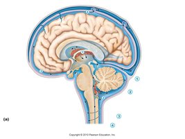
CSF is produced by choroid plexuses in the ventricles of the brain. It circulates through the ventricles, into the subarachnoid space, and is absorbed into the dural venous sinuses via arachnoid granulations.
Normal volume: ~150 mL, replaced every 8 hours.
Choroid plexuses filter blood plasma and remove wastes.
Clinical Correlate: Hydrocephalus
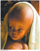
Hydrocephalus is the abnormal accumulation of CSF within the brain's ventricles, leading to increased intracranial pressure and potential brain damage.
Blood-Brain Barrier (BBB)
Structure and Function
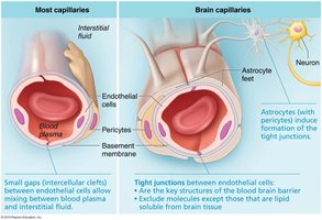
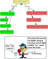
The blood-brain barrier maintains a stable environment for the brain by restricting the passage of harmful substances. It is composed of:
Continuous endothelium of capillary walls with tight junctions
Thick basal lamina containing enzymes that degrade certain chemicals
Astrocyte feet and pericytes that support and regulate the barrier
Permeability and Exceptions
The BBB is not uniform throughout the brain. It is more permeable in regions such as the choroid plexuses, the vomiting center, and the hypothalamus. Substances that can cross include glucose, essential amino acids, some electrolytes (via facilitated diffusion), and fat-soluble molecules (e.g., alcohol, nicotine, anesthetics).
The Spinal Cord
Gross Anatomy and Protection
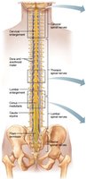
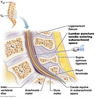
The spinal cord is enclosed within the vertebral column, extending from the foramen magnum to the level of L1 or L2. It is anchored by the filum terminale and denticulate ligaments (extensions of pia mater). The spinal cord features cervical and lumbar enlargements for limb innervation and terminates in the conus medullaris. The cauda equina is a bundle of nerve roots at the base of the spine.
Cross-Sectional Anatomy
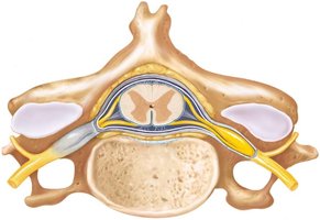
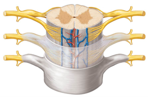
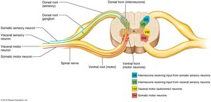
The spinal cord is partially divided by the ventral median fissure and dorsal median sulcus. Gray matter is organized into anterior (ventral) and posterior (dorsal) horns, with lateral horns present in thoracic and superior lumbar regions. The gray commissure connects the two sides and encloses the central canal.
Spinal Roots and Nerves
Spinal nerves are formed by the fusion of dorsal (sensory) and ventral (motor) roots. The dorsal and ventral roots are part of the PNS, not the CNS.
White Matter and Spinal Tracts
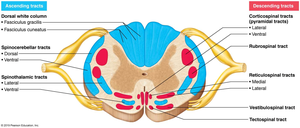
White matter consists of myelinated and unmyelinated fibers that allow communication between the spinal cord and brain. Fibers run in three directions: ascending (sensory), descending (motor), and transverse (commissural). White matter is organized into dorsal, lateral, and ventral columns (funiculi), each containing specific tracts.
Neuronal Pathways
Spinal tracts exhibit four general properties:
Decussation: Most tracts cross from one side of the CNS to the other.
Relay: Pathways consist of two or three neurons in sequence.
Somatotopy: Spatial arrangement reflects the body region served.
Symmetry: Pathways are paired on both sides of the CNS.
Peripheral Nervous System (PNS)
Components and Structure of a Nerve
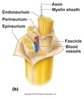
The PNS includes all neural structures outside the brain and spinal cord: sensory receptors, peripheral nerves, ganglia, and motor endings. A nerve is a cordlike organ composed of bundles of axons (myelinated and unmyelinated) surrounded by connective tissue layers:
Endoneurium: Surrounds individual axons
Perineurium: Bundles axons into fascicles
Epineurium: Encloses the entire nerve
Cranial Nerves
Overview and Classification
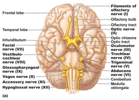
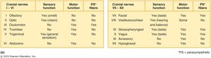
There are 12 pairs of cranial nerves, each with specific sensory, motor, or mixed functions. Most serve the head and neck, except the vagus nerve, which extends into the thorax and abdomen. Cranial nerves are numbered I–XII from rostral to caudal.
Selected Cranial Nerves
Olfactory (I): Sensory for smell
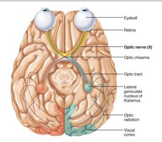
Optic (II): Sensory for vision; fibers cross at the optic chiasma for binocular vision
Oculomotor (III), Trochlear (IV), Abducens (VI): Motor to extrinsic eye muscles
Trigeminal (V): Mixed; sensory from face, motor for mastication
Facial (VII): Mixed; motor for facial expression, sensory for taste
Vestibulocochlear (VIII): Sensory for hearing and balance
Glossopharyngeal (IX), Vagus (X), Accessory (XI), Hypoglossal (XII): Mixed or motor; functions include swallowing, taste, parasympathetic output, and head/neck movement
Spinal Nerves and Plexuses
Spinal Nerve Organization
There are 31 pairs of mixed spinal nerves, named for their region of origin: cervical (C1–C8), thoracic (T1–T12), lumbar (L1–L5), sacral (S1–S5), and coccygeal (C0). Each spinal nerve branches into dorsal and ventral rami, which carry both sensory and motor fibers.
Nerve Plexuses
Except for T2–T12, ventral rami form plexuses (cervical, brachial, lumbar, sacral) that redistribute fibers so each limb muscle receives input from more than one spinal nerve. This redundancy protects against complete paralysis from single nerve injury.
Dermatomes
A dermatome is an area of skin innervated by the sensory fibers of a single spinal nerve. All spinal nerves except C1 participate in dermatomes, which are clinically important for diagnosing nerve or spinal cord injuries.
