 Back
BackPulmonary Ventilation and Gas Exchange: Mechanisms and Regulation
Study Guide - Smart Notes
 Tailored notes based on your materials, expanded with key definitions, examples, and context.
Pulmonary Ventilation and Gas Exchange
Overview of Respiration
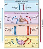
Respiration is the process by which gases are exchanged between the atmosphere and the body’s cells. It involves four main processes: pulmonary ventilation, pulmonary gas exchange, gas transport, and tissue gas exchange. Each step is essential for the delivery of oxygen to tissues and the removal of carbon dioxide from the body.
Pulmonary ventilation: Movement of air between the atmosphere and alveoli.
Pulmonary gas exchange: Exchange of gases between alveoli and blood.
Gas transport: Transport of gases in the blood between lungs and systemic cells.
Tissue gas exchange: Exchange of gases between blood and systemic cells.
Mechanics of Breathing
Muscles Involved in Breathing
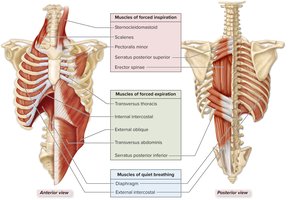
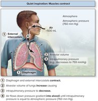
Breathing is accomplished by the coordinated action of several skeletal muscles. The diaphragm and external intercostals are the primary muscles for quiet breathing, while additional muscles are recruited during forced inspiration and expiration.
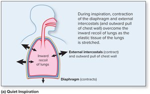
Quiet inspiration: Diaphragm contracts and flattens; external intercostals elevate the ribs.
Forced inspiration: Sternocleidomastoid, scalenes, pectoralis minor, serratus posterior superior, and erector spinae further increase thoracic volume.
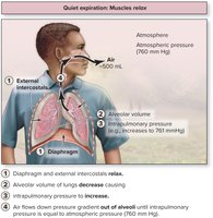
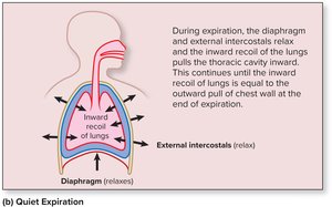
Forced expiration: Internal intercostals, abdominal muscles, transversus thoracis, and serratus posterior inferior contract to decrease thoracic volume.
Thoracic Cavity Volume Changes
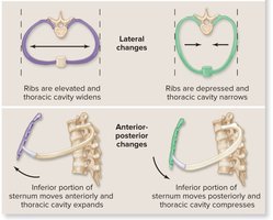
Changes in the dimensions of the thoracic cavity drive the movement of air into and out of the lungs. These changes occur in three planes: vertical, lateral, and anterior-posterior.
Vertical changes: Diaphragm contraction increases vertical dimension; relaxation decreases it.
Lateral changes: Elevation of ribs widens the thoracic cavity; depression narrows it.
Anterior-posterior changes: Sternum moves anteriorly during inspiration and posteriorly during expiration.
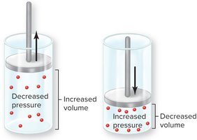
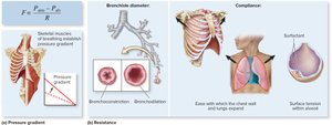
Boyle’s Law and Pressure Gradients
Boyle’s Law states that at a constant temperature, the pressure of a gas is inversely related to its volume. This principle explains how changes in thoracic volume create pressure gradients that drive airflow.
Equation:
Increasing thoracic volume decreases intrapulmonary pressure, causing air to flow in (inspiration).
Decreasing thoracic volume increases intrapulmonary pressure, causing air to flow out (expiration).
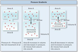
Pressure Gradients and Airflow
Air moves from regions of higher pressure to lower pressure. During inspiration, the pressure inside the lungs becomes lower than atmospheric pressure, drawing air in. During expiration, the pressure inside the lungs exceeds atmospheric pressure, pushing air out.
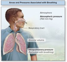
Volumes and Pressures Associated with Breathing
Several pressures are important in breathing:
Atmospheric pressure: Pressure of air in the environment (760 mm Hg at sea level).
Intrapulmonary pressure: Pressure within the alveoli; fluctuates with breathing.
Intrapleural pressure: Pressure within the pleural cavity; always lower than intrapulmonary pressure to keep lungs inflated.
Mechanics of Quiet Breathing
During quiet inspiration, the diaphragm and external intercostals contract, increasing thoracic volume and decreasing intrapulmonary pressure, allowing air to flow in. During quiet expiration, these muscles relax, thoracic volume decreases, and air is expelled as pressure increases.
Nervous Control of Breathing
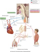
Respiratory Centers in the Brainstem
Breathing is regulated by autonomic nuclei in the brainstem, specifically the medullary respiratory center (ventral and dorsal respiratory groups) and the pontine respiratory center. These centers coordinate the rhythmic contraction and relaxation of respiratory muscles.
VRG (Ventral Respiratory Group): Controls motor output to respiratory muscles.
DRG (Dorsal Respiratory Group): Integrates sensory input and modifies VRG activity.
Pontine center: Smooths transitions between inspiration and expiration.

Airflow, Pressure Gradients, and Resistance
Determinants of Airflow
Airflow is determined by the pressure gradient between the atmosphere and the alveoli, and by resistance within the airways. The relationship is described by the equation:
Equation:
F: Airflow
ΔP: Pressure gradient
R: Resistance
Measuring Respiratory Function
Respiratory Volumes and Capacities
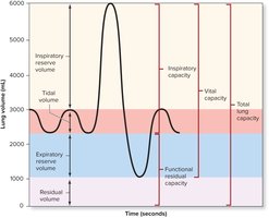
Spirometry is used to measure lung volumes and capacities, which are important for assessing respiratory health.
Tidal volume (TV): Air inhaled or exhaled per breath during quiet breathing.
Inspiratory reserve volume (IRV): Additional air that can be inhaled after a normal inspiration.
Expiratory reserve volume (ERV): Additional air that can be exhaled after a normal expiration.
Residual volume (RV): Air remaining in lungs after maximal expiration.
Vital capacity (VC): TV + IRV + ERV.
Total lung capacity (TLC): TV + IRV + ERV + RV.
Volume/Capacity | Definition | Typical Value (Male) | Typical Value (Female) |
|---|---|---|---|
Tidal Volume (TV) | Amount of air taken in or expelled during quiet breath | 500 mL | 500 mL |
Inspiratory Reserve Volume (IRV) | Amount forcibly inhaled beyond tidal volume | 3100 mL | 1900 mL |
Expiratory Reserve Volume (ERV) | Amount forcibly exhaled beyond tidal volume | 1200 mL | 700 mL |
Residual Volume (RV) | Amount left in lungs after forced expiration | 1200 mL | 1100 mL |
Vital Capacity (VC) | TV + IRV + ERV | 4800 mL | 3100 mL |
Total Lung Capacity (TLC) | TV + IRV + ERV + RV | 6000 mL | 4200 mL |
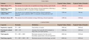
Additional info: These values can vary with age, sex, body size, and physical conditioning.
