 Back
BackRespiratory Part 3 AP 2nd year Thoracic cavity spirometry
Study Guide - Smart Notes
 Tailored notes based on your materials, expanded with key definitions, examples, and context.
Pressure Relationships in the Thoracic Cavity
Intrapulmonary and Intrapleural Pressures
The thoracic cavity contains several important pressure relationships that are essential for normal breathing. These include:
Intrapulmonary Pressure (Ppul): The pressure within the alveoli, also called intra-alveolar pressure. It fluctuates with the phases of breathing and always eventually equalizes with atmospheric pressure (Patm).
Intrapleural Pressure (Pip): The pressure within the pleural cavity. It is always negative relative to both atmospheric and intrapulmonary pressures, typically about 4 mmHg less than Ppul. This negative pressure is crucial for keeping the lungs inflated.
Transpulmonary Pressure: The difference between intrapulmonary and intrapleural pressures. It keeps the airways open and prevents lung collapse.
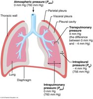
Clinical Note: If intrapleural pressure becomes positive (e.g., due to fluid accumulation), lung collapse can occur. Each lung is in a separate cavity, so one can collapse without affecting the other.
Pulmonary Ventilation
Mechanics of Breathing
Pulmonary ventilation is the mechanical process of moving air into and out of the lungs. It depends on volume changes in the thoracic cavity, which lead to pressure changes and the movement of gases to equalize pressure.
Boyle’s Law and Its Application
Boyle’s Law describes the inverse relationship between the pressure and volume of a gas at constant temperature:
As the volume of a container increases, the pressure decreases, and vice versa.
Mathematically:
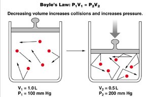
This principle explains how changes in thoracic volume during breathing alter lung pressures and drive airflow.
Inspiration
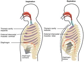
Inspiration is an active process involving the contraction of the diaphragm and external intercostal muscles:
Diaphragm: Contracts and moves downward, increasing thoracic volume.
External Intercostals: Contract to lift the rib cage up and out, further increasing thoracic volume (like raising a bucket handle).
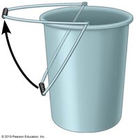
As thoracic volume increases, intrapulmonary pressure drops below atmospheric pressure, causing air to flow into the lungs until equilibrium is reached.
Forced Inspiration
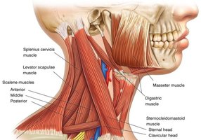
During exercise or respiratory distress, additional muscles (scalenes, sternocleidomastoid, pectoralis minor) are recruited to further increase thoracic volume and create a larger pressure gradient for airflow.
Expiration
Expiration is usually a passive process during quiet breathing, resulting from relaxation of inspiratory muscles and elastic recoil of the lungs. Forced expiration is active and involves abdominal and internal intercostal muscles.
Factors Influencing Pulmonary Ventilation
Airway Resistance
Airway resistance is primarily due to friction in the airways. The relationship is described by:
, where F is airflow, ΔP is the pressure gradient, and R is resistance.
Resistance is usually low due to large airway diameters and extensive branching, which increases total cross-sectional area.
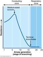
Severe constriction (e.g., asthma) increases resistance and can stop ventilation. Epinephrine can dilate bronchioles and reduce resistance.

Alveolar Surface Tension
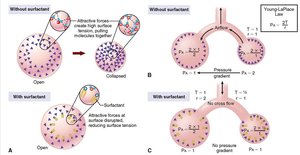
Surface tension arises from the attraction between liquid molecules at the alveolar surface, which can impede lung expansion. Surfactant reduces this tension, making it easier to inflate the lungs.
Lung Compliance
Lung compliance is a measure of the lung's ability to stretch and expand. High compliance means the lungs expand easily, aided by surfactant and the elasticity of lung tissue.
Ventilation Assessment
Spirometry
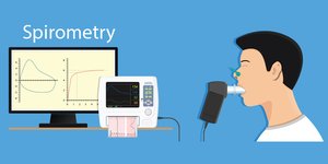
Spirometry is used to measure respiratory volumes and capacities, providing valuable information about pulmonary function. Abnormal results can indicate respiratory disorders.
Lung Volumes and Capacities
Key respiratory volumes include tidal volume, inspiratory reserve volume, expiratory reserve volume, and residual volume. These combine to form capacities such as vital capacity and total lung capacity.
Alveolar Ventilation
Minute and Alveolar Ventilation
Minute ventilation is the total volume of gas entering or leaving the respiratory tract per minute. Alveolar ventilation rate (AVR) is a better indicator of effective ventilation, as it measures the flow of gases into and out of the alveoli.
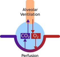
The formula for alveolar ventilation is:
Alveolar ventilation = BF × (TV – dead space)
BF = breathing frequency (breaths/min)
TV = tidal volume (ml/breath)
Dead space is the volume of airways where no gas exchange occurs.
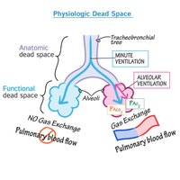
Since dead space is relatively constant, changes in alveolar ventilation are primarily due to changes in breathing frequency and tidal volume.
