 Back
BackPulmonary Ventilation and the Respiratory System: Structure, Function, and Clinical Significance
Study Guide - Smart Notes
 Tailored notes based on your materials, expanded with key definitions, examples, and context.
The Respiratory System: Pulmonary Ventilation
Overview of Respiratory Function
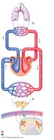
The respiratory system is essential for gas exchange, supplying oxygen to the body and removing carbon dioxide. It supports both external respiration (gas exchange between the atmosphere and blood) and internal respiration (gas exchange between blood and tissues). The process of pulmonary ventilation refers to the movement of air into and out of the lungs, driven by pressure gradients.
Internal respiration: Cellular utilization of oxygen and production of ATP via oxidative phosphorylation.
External respiration: Includes pulmonary ventilation, exchange of gases between lungs and blood, transport of gases in blood, and exchange between blood and tissues.
Other functions: Acid-base balance, vocalization, defense against pathogens, water and heat loss, enhancing venous return, and activation of plasma proteins.
Anatomy of the Respiratory System
Upper Airways and Respiratory Tract
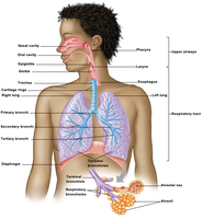
The respiratory system is divided into the upper airways and the respiratory tract. The upper airways include the nasal and oral cavities and the pharynx. The respiratory tract extends from the larynx to the lungs and is divided into the conducting and respiratory zones.
Upper airways: Nasal cavity, oral cavity, pharynx.
Respiratory tract: Larynx, trachea, bronchi, bronchioles, alveoli.
Conducting zone: Air passageways that do not participate in gas exchange (from larynx to terminal bronchioles).
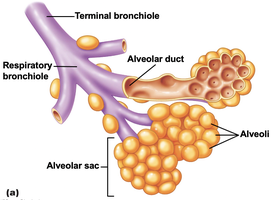
Respiratory zone: Sites of gas exchange (respiratory bronchioles, alveolar ducts, alveoli).
The Conducting Zone
The conducting zone includes the larynx, trachea, bronchi, and bronchioles. It serves as a passageway for air, warms and humidifies incoming air, and filters particles via mucus and cilia.
Larynx: Contains the glottis and epiglottis, which protect the airway during swallowing.
Trachea: Supported by C-shaped cartilage rings for rigidity.
Bronchi: Primary, secondary, and tertiary branches supply each lung lobe.
Bronchioles: Smallest airways, less than 1 mm in diameter.
Epithelium: Goblet cells secrete mucus; ciliated cells move mucus toward the mouth (mucus escalator).
The Respiratory Zone
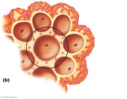
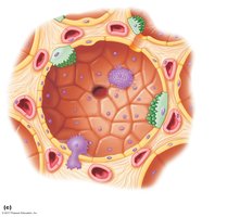
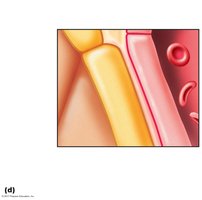
The respiratory zone is where gas exchange occurs by diffusion. It includes respiratory bronchioles, alveolar ducts, and alveoli. Alveoli are the primary site of gas exchange and are surrounded by a rich capillary network.
Alveoli: Approximately 300 million in the lungs, providing a large surface area for gas exchange.
Type I alveolar cells: Form the alveolar wall (single layer of epithelial cells).
Type II alveolar cells: Secrete surfactant to reduce surface tension.
Alveolar macrophages: Provide immune defense.
Thoracic Cavity and Pleura
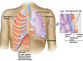
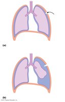
The thoracic cavity houses the lungs and is protected by the chest wall (ribs, sternum, vertebrae, and muscles). The pleura are double-layered membranes that surround each lung and line the chest wall, with a small amount of intrapleural fluid in between to reduce friction and maintain lung expansion.
Pleural sac: Encloses each lung, creating the intrapleural space.
Intrapleural fluid: Lubricates and helps keep the lungs expanded.
Forces for Pulmonary Ventilation
Pressure Gradients and Airflow
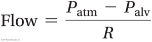
Air moves in and out of the lungs by bulk flow, driven by pressure gradients. Air flows from regions of higher to lower pressure. The main pressures involved are atmospheric pressure (Patm), intra-alveolar pressure (Palv), intrapleural pressure (Pip), and transpulmonary pressure (Palv – Pip).
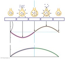
Inspiration: Palv < Patm (air flows into lungs).
Expiration: Palv > Patm (air flows out of lungs).
Transpulmonary pressure: The difference between Palv and Pip; determines lung expansion.
Pulmonary Pressures
Atmospheric pressure (Patm): Pressure of air outside the body (760 mm Hg at sea level).
Intra-alveolar pressure (Palv): Pressure within the alveoli; varies with breathing cycle.
Intrapleural pressure (Pip): Pressure within the pleural sac; always negative under normal conditions.
Transpulmonary pressure:
Mechanics of Breathing
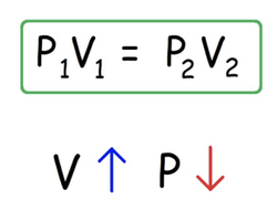
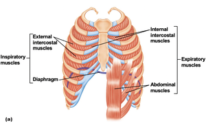
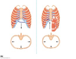
Changes in thoracic cavity volume alter alveolar pressure, driving ventilation. The primary muscles involved are the diaphragm and intercostal muscles.
Inspiration: Diaphragm and external intercostals contract, increasing thoracic volume and decreasing Palv.
Expiration: Usually passive; internal intercostals and abdominal muscles contract during active expiration.
Factors Affecting Pulmonary Ventilation
Lung Compliance
Lung compliance refers to the ease with which the lungs can be stretched. High compliance means the lungs expand easily; low compliance means more effort is required.
Elasticity: More elastic tissue decreases compliance.
Surface tension: Created by water molecules lining the alveoli; surfactant reduces surface tension and increases compliance.
Airway Resistance
Airway resistance is influenced by airway diameter, smooth muscle tone, and mucus. As airways branch and become smaller, their total cross-sectional area increases, keeping overall resistance low.
Bronchoconstriction: Decreases airway radius, increasing resistance (mediated by parasympathetic activity, histamine).
Bronchodilation: Increases airway radius, decreasing resistance (mediated by sympathetic activity, epinephrine, CO2).
Clinical Significance of Respiratory Volumes and Air Flows
Lung Volumes and Capacities
Lung volumes and capacities are measured to assess pulmonary function. Spirometry is a common method for measuring these values.
Volume/Capacity | Definition | Normal Value (mL) |
|---|---|---|
Tidal Volume (VT) | Volume of air in a single, unforced breath | 500 |
Inspiratory Reserve Volume (IRV) | Volume that can be inspired after normal inspiration | 3000 |
Expiratory Reserve Volume (ERV) | Volume that can be expired after normal expiration | 1000 |
Residual Volume (RV) | Volume remaining after maximal expiration | 1200 |
Inspiratory Capacity (IC) | VT + IRV | 3500 |
Vital Capacity (VC) | VT + IRV + ERV | 4500 |
Functional Residual Capacity (FRC) | ERV + RV | 2200 |
Total Lung Capacity (TLC) | VT + IRV + ERV + RV | 5700 |
Pulmonary Function Tests
Obstructive diseases: Increased airway resistance (e.g., asthma, COPD); increased RV and FRC, decreased VC.
Restrictive diseases: Decreased lung expansion (e.g., fibrosis); decreased TLC and VC.
Forced Vital Capacity (FVC): Maximum volume exhaled after maximum inhalation.
Forced Expiratory Volume (FEV1): Percentage of FVC exhaled in 1 second; normal is 80%.
Peak Expiratory Flow Rate (PEFR): Maximum rate of exhalation; men ~9 L/sec, women ~7 L/sec.
Alveolar Ventilation
Alveolar ventilation is the volume of fresh air reaching the alveoli per minute. It is more accurate than minute ventilation for assessing effective gas exchange.
Minute ventilation:
Anatomical dead space: Volume of air in conducting zone (~150 mL) that does not participate in gas exchange.
Key Definitions
Tidal volume (VT): Volume of air moved in a single, unforced breath.
Minute ventilation: Total volume of air entering/leaving the respiratory system per minute.
Intra-alveolar pressure (Palv): Pressure of air within the alveoli.
Anatomical dead space: Air in the conducting zone not involved in gas exchange.
Alveolar ventilation: Volume of fresh air reaching the alveoli per minute.
Sample Study Questions
Describe the differences between the conducting zone and the respiratory zone.
Which structures make up the respiratory membrane?
During inspiration, which is greater: intra-alveolar pressure or atmospheric pressure?
Which muscles contribute to inspiration? Expiration?
Describe two instances that illustrate the importance of recoil in the respiratory system.
If scarring of the lung resulted from some disease process, what would happen to lung compliance?
If surfactant secretion decreased, what would happen to lung compliance?
Will contraction of bronchiole smooth muscle cause the resistance to air flow to increase or decrease?
Following a maximum inspiration, a person expires maximally. Which lung capacity corresponds to the volume of air that has been expired? What is the volume of air left in the lungs called?
