 Back
BackPulmonary Ventilation: Mechanics, Physical Factors, and Pulmonary Volumes 21.3
Study Guide - Smart Notes
 Tailored notes based on your materials, expanded with key definitions, examples, and context.
Pulmonary Ventilation
Introduction to Pulmonary Ventilation
Pulmonary ventilation, or breathing, is the process of moving air into and out of the lungs.
consists of two main phases: inspiration (inhalation) and expiration (exhalation). The movement of air is driven by pressure gradients, which are created by changes in the volume of the thoracic cavity and lungs.
The Pressure–Volume Relationship in Pulmonary Ventilation
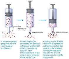
Gas molecules move from areas of higher pressure to areas of lower pressure.
Boyle’s Law and Its Application
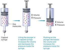
Boyle’s law states that at a constant temperature and number of gas molecules, the pressure and volume of a gas are inversely related. This principle is fundamental to understanding how air moves in and out of the lungs during breathing.
As volume increases, pressure decreases.
As volume decreases, pressure increases.
In the context of the lungs, increasing the thoracic volume during inspiration decreases intrapulmonary pressure, allowing air to flow in. Conversely, decreasing thoracic volume during expiration increases intrapulmonary pressure, pushing air out.
Pressure Gradients and Airflow
Air moves from regions of higher pressure to regions of lower pressure. During inspiration, the pressure inside the lungs becomes lower than atmospheric pressure, causing air to flow in. During expiration, the pressure inside the lungs exceeds atmospheric pressure, causing air to flow out.
Pressure Changes During Pulmonary Ventilation
Types of Pressures Involved
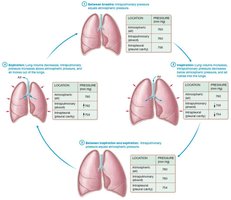
Atmospheric Pressure: The pressure exerted by the air surrounding the body, typically 760 mm Hg at sea level.
It increases as you go below sea level, and it decreases as you rise above sea level.
Intrapulmonary Pressure: The air pressure within the alveoli, which rises and falls with breathing but always equalizes with atmospheric pressure due to pressure gradients reaching equilibrium.
Intrapleural Pressure: The pressure within the pleural cavity, normally about 4 mm Hg less than intrapulmonary pressure, preventing lung collapse.
Intrapleural pressure also rises and falls with inspiration and expiration but does not equalize with atmospheric pressure.
Pressure Changes During the Breathing Cycle
During the breathing cycle, the following steps occur:
At rest: Intrapulmonary pressure equals atmospheric pressure; no air movement.
Inspiration: Thoracic volume increases, intrapulmonary pressure drops below atmospheric, and air flows in.
End of inspiration: Intrapulmonary pressure equals atmospheric pressure; airflow stops.
Expiration: Thoracic volume decreases, intrapulmonary pressure rises above atmospheric, and air flows out.
Lung Collapse and Pneumothorax
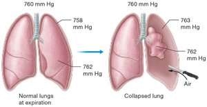
If intrapleural pressure rises to atmospheric or above, the suction effect is lost, and the lung collapses. This can occur due to trauma, pleural effusion, pneumothorax, or hemothorax.
When the lung is fully collapsed, intrapulmonary pressure is so high that the body is unable to bring it below atmospheric pressure, and inspiration cannot occur
Mechanics of Inspiration and Expiration
Muscles Involved in Ventilation
The lungs rely on skeletal muscles of the thoracic cavity to change/ increase their volume.
The Inspiratory muscles--they increase the size of the thoracic cavity, which indirectly increases the volume of the lungs.
When the thoracic cavity increases in height and diameter, the parietal pleura is pulled with it. The parietal pleura, in turn, pulls on the visceral pleura, which pulls the lungs outward, increasing their volume.
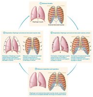
The main inspiratory muscle is the diaphragm, assisted by the external intercostals (located between the ribs).
When relaxed between breaths, the diaphragm muscle is dome-shaped and bulges up into the thoracic cavity. This keeps the volume of the lungs low.
When the diaphragm muscle contracts, it moves from its relaxed dome shape and pulls down to become flat. This action increases the height of the thoracic cavity.
Both actions increase the volume of the lungs, which decreases the intrapulmonary pressure
The diaphragm and external intercostal muscles remain contracted to hold the lungs at the increased volume.
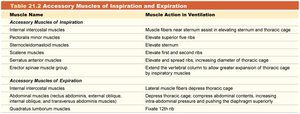
During deep or forced inspiration, accessory muscles such as the internal intercostals, pectoralis minor,
sternocleidomastoid, scalene, serratus anterior, and certain back muscles are also involved.
The resulting additional increase in lung volume further decreases intrapulmonary pressure, moving air into the lungs.
When the inspiratory muscles relax, two things happen: (1) the diaphragm resumes its original dome shape, which pushes up on the lungs; and (2) the elastic tissue in the lungs recoils, and the lungs snap back to a smaller size.
These two actions decrease lung volume and raise intrapulmonary pressure above atmospheric pressure, so air flows out of the lungs.
When expiration is forced, the accessory muscles of expiration come into play. These muscles, which include the internal intercostal, abdominal, and certain back muscles, forcefully decrease the size of the thoracic cavity.
Expiration may also be forced by other means, such as slapping a person on the back or delivering abdominal thrusts that push up on the diaphragm muscle (commonly called the Heimlich maneuver.
Accessory Muscles of Inspiration and Expiration
Muscle Name | Muscle Action in Ventilation |
|---|---|
Internal intercostal muscles | Assist in elevating sternum and thoracic cage |
Pectoralis minor muscles | Elevate superior ribs |
Sternocleidomastoid muscles | Elevate sternum |
Scalene muscles | Elevate first and second ribs |
Serratus anterior muscles | Elevate and spread ribs, increasing diameter of thoracic cage |
Erector spinae muscle group | Extend the vertebral column to allow greater expansion of thoracic cage by inspiratory muscles |
Abdominal muscles, certain back muscles | Forcefully decrease thoracic cavity size during expiration |
Nonrespiratory Movements
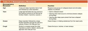
Movements such as yawns, coughs, sighs, and sneezes are not intended for ventilation but serve other functions, such as clearing airways or reopening collapsed alveoli.
Movement | Definition | Function |
|---|---|---|
Sigh | A slow and deep inspiration that is held and followed by a slow expiration | Reopens local groups of collapsed alveoli and stimulates release of surfactant |
Yawn | Large sigh that takes lung volume to inspiratory capacity | Opens collapsed alveoli, minimizes alveolar collapse during sleep |
Sneeze | Deep inspiration followed by a large, forceful expiration through the nose | Clears foreign or irritating substances from the nasal cavity |
Cough | Small or absent initial inspiration, followed by forceful expiration | Clears the larynx, trachea, or lower airways |
PUTTING IT ALL TOGETHER:
Between breaths, the intrapulmonary and atmospheric pressures are equal, so no air moves between them.
During inspiration, the inspiratory muscles contract, which increases the volume of the lungs. This decreases intrapulmonary pressure below atmospheric pressure, and air flows into the lungs.
Between inspiration and expiration, intrapulmonary pressure is again equal to atmospheric pressure and no air movement occurs.
During expiration, the lungs’ volume decreases because the inspiratory muscles relax and the lungs’ elastic tissue recoils. This increases intrapulmonary pressure above atmospheric pressure, and air flows out of the lungs.
Physical Factors Influencing Pulmonary Ventilation
The three primary physical factors are airway resistance, alveolar surface tension, and pulmonary compliance
Airway Resistance
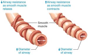
Airway resistance is anything that impedes airflow through the respiratory tract. It is primarily determined by the diameter of the airways, which can be altered by smooth muscle contraction (bronchoconstriction) or relaxation (bronchodilation)
Resistance is determined by diameter.
Resistance normally varies during pulmonary ventilation due to changes in intrapulmonary pressure.
During inspiration, resistance decreases slightly because the airways are pulled open as the lungs expand.
During expiration, resistance increases slightly as the lungs recoil and the airways narrow.
The diameter of the airways is also controlled by the contraction or relaxation of the smooth muscle of the bronchioles.
Relaxation of the bronchial smooth muscle, or bronchodilation, increases the diameter of the bronchioles
Neurons of the sympathetic nervous system release norepinephrine during times of exercise, stress, or emergency, which triggers bronchodilation to increase the efficiency of pulmonary ventilation.
Conversely, when the smooth muscle contracts, a process called bronchoconstriction, the diameter of the bronchioles can decrease.
This narrows the lumen of the bronchiole, which dramatically increases resistance.
Bronchoconstriction may be triggered by inhaled irritants that activate the parasympathetic neurons serving the bronchial smooth muscle or by inflammatory mediators
Certain disease states may also increase airway resistance by causing inflammation or causing an obstruction such as a tumor or excess mucus. (ex. COVID)
Alveolar Surface Tension
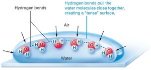
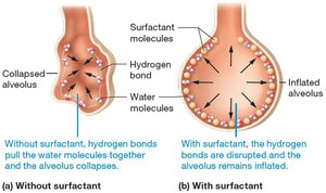
Alveoli are lined with a thin film of water, creating a gas–water boundary. Water molecules form hydrogen bonds, generating surface tension that tends to collapse the alveolus, especially during expiration. Surfactant, produced by Type II alveolar cells, reduces this surface tension and prevents alveolar collapse (atelectasis).
When surfactant is added to water, its polar ends interact with water molecules while its nonpolar ends repel water molecules. This action disrupts hydrogen bonds between water molecules, which reduces alveolar surface tension. The reduced surface tension allows the alveoli to remain partially open, even during expiration
Pulmonary Compliance
Pulmonary compliance refers to the ability of the lungs and chest wall to stretch (a property known as distensibility). It is influenced by alveolar surface tension, the distensibility of lung tissue, and the mobility of the chest wall. Decreased compliance makes lung expansion more difficult and reduces the effectiveness of ventilation.
Pulmonary compliance is primarily determined by three factors:
Degree of alveolar surface tension. Surface tension on the alveoli resists their inflation, but surfactant counters this tension.
Distensibility of elastic tissue in the lungs. The elastic tissue in the lungs gives them their ability to stretch during inflation and to recoil during expiration.
Ability of the chest wall to move. The chest wall must also stretch during inspiration, as the lungs and chest wall move together (mobility of chest wall).
Factors that decrease pulmonary compliance make pulmonary ventilation less efficient and increase the work of breathing.
For example, diseases that decrease surfactant production can result in alveolar surface tension that is too high, which makes the alveoli difficult to inflate during inspiration
Anything that damages the lung, including infections such as tuberculosis or foreign particles such as coal dust, can destroy the lungs’ elastic tissue.
In these situations, the destroyed elastic tissue is replaced with dense irregular connective tissue, producing a state called fibrosis . This stiffens the lungs, decreasing their compliance and making them difficult to inflate.
Pulmonary Volumes and Capacities
Measuring Pulmonary Volumes
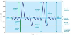
Pulmonary volumes are measured using a spirometer (A laboratory instrument that measures volumes of air exchanged with ventilation) and are useful for assessing lung function. The main volumes include:
Tidal Volume (TV): Amount of air inspired or expired during normal quiet breathing (~500 ml).
If we multiply the TV by the number of breaths per minute, we get the minute volume, the total volume of air that moves in and out of the lungs each minute.
The air that remains in the conducting zone airways is said to be in the anatomical dead space.
Alveolar ventilation rate (AVR), the volume of air that reaches the alveoli multiplied by the breaths per minute
The AVR averages about 4.2 liters per minute.
Inspiratory Reserve Volume (IRV): Volume that can be forcibly inspired after a normal inspiration (2100–3300 ml depending on a persons sex and size).
Expiratory Reserve Volume (ERV): Volume that can be forcibly expired after a normal expiration (700–1200 ml). (IRV in reverse)
Residual Volume (RV): Air remaining in lungs after forceful expiration.
The RV remains in the lungs due mostly to the intrapleural pressure and outward recoil of the chest wall, which keep the lungs slightly inflated.
Pulmonary Capacities
Inspiratory Capacity: TV (tidal volume) + IRV (Inspiratory Reserve Volume)
The total amount of air that a person can inspire after a tidal expiration.
Functional Residual Capacity: ERV (Expiratory Reserve Volume) + RV (Residual Volume)
Is the amount of air that is normally left in the lungs after a tidal expiration.
Vital Capacity: TV (Tidal Volume) + IRV (Inspiratory Reserve Volume) + ERV (Expiratory Reserve Volume)
Represents the total amount of exchangeable air, or the total amount of air that can move in and out of the lungs.
The vital capacity is measured in the laboratory with a spirometer as the forced vital capacity (FVC), in which the subject follows a maximal inspiration with a maximal exhalation.
Total Lung Capacity (TLC): IRV (Inspiratory Reserve Volume) + TV (Tidal Volume) + ERV (Expiratory Reserve Volume) + RV (Residual Volume)
Represents the total amount of exchangeable and nonexchangeable air in the lungs.
Summary Table of Pulmonary Volumes and Capacities
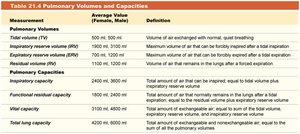
Measurement | Average Value (Female, Male) | Definition |
|---|---|---|
Tidal volume (TV) | 500 ml, 500 ml | Volume of air exchanged with normal, quiet breathing |
Inspiratory reserve volume (IRV) | 1900 ml, 3100 ml | Maximum volume of air that can be forcibly inspired after a tidal inspiration |
Expiratory reserve volume (ERV) | 700 ml, 1200 ml | Maximum volume of air that can be forcibly expired after a tidal expiration |
Residual volume (RV) | 1100 ml, 1200 ml | Volume of air that remains in the lungs after a forced expiration |
Inspiratory capacity | 2400 ml, 3600 ml | Total amount of air that can be inspired, equal to tidal volume plus inspiratory reserve volume |
Functional residual capacity | 1800 ml, 2400 ml | Amount of air that normally remains in the lungs after a tidal expiration, equal to the residual volume plus expiratory reserve volume |
Vital capacity | 3100 ml, 4800 ml | Total amount of exchangeable air, or the total amount of air that can move in and out of the lungs |
Total lung capacity | 4200 ml, 6000 ml | Total amount of exchangeable and nonexchangeable air, equal to the sum of all the pulmonary volumes |
Additional info: Pulmonary volumes and capacities are influenced by factors such as age, sex, body size, and health status. Values are generally lower in females than in males and decrease with age or certain diseases.
