 Back
BackPulmonary Ventilation: Mechanisms and Physical Factors
Study Guide - Smart Notes
 Tailored notes based on your materials, expanded with key definitions, examples, and context.
Pulmonary Ventilation
Introduction to Pulmonary Ventilation
Pulmonary ventilation is the process of moving air into and out of the lungs, enabling gas exchange with the environment. It consists of two main phases: inspiration (inhaling air into the lungs) and expiration (exhaling air out of the lungs).
Pressure-Volume Relationships in Pulmonary Ventilation
Boyle’s Law and Pulmonary Mechanics
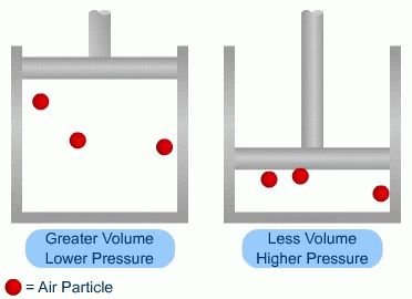
Boyle’s law states that at a constant temperature and number of gas molecules, the pressure and volume of a gas are inversely related. This principle is fundamental to understanding how air moves in and out of the lungs:
As lung volume increases, intrapulmonary pressure decreases, causing air to flow into the lungs.
As lung volume decreases, intrapulmonary pressure increases, causing air to flow out of the lungs.
Gas always moves from areas of higher pressure to areas of lower pressure.
Pressure Gradients and Pulmonary Ventilation
Types of Pressures Involved
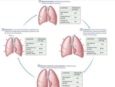
Atmospheric Pressure: The pressure exerted by air in the environment (at sea level, 760 mmHg).
Intrapulmonary Pressure: The pressure within the alveoli, fluctuates with breathing and eventually equilibrates with atmospheric pressure.
Intrapleural Pressure: The pressure within the pleural cavity, always slightly less than intrapulmonary pressure (about 4 mmHg lower), preventing lung collapse.
Cycle of Pulmonary Ventilation
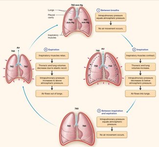
At rest: Intrapulmonary pressure equals atmospheric pressure; no air movement.
Inspiration: Intrapulmonary pressure drops below atmospheric pressure; air flows into lungs.
Between inspiration and expiration: Pressures equalize; no air movement.
Expiration: Intrapulmonary pressure rises above atmospheric pressure; air flows out of lungs.
Mechanics of Inspiration and Expiration
Muscles Involved in Ventilation
The lungs themselves cannot change volume; they rely on skeletal muscles to alter thoracic cavity size:
Main inspiratory muscles: Diaphragm and external intercostal muscles.
Accessory muscles: Used during forced inspiration or expiration.
If the diaphragm is damaged, inspiration is weakened and the body relies on accessory muscles.
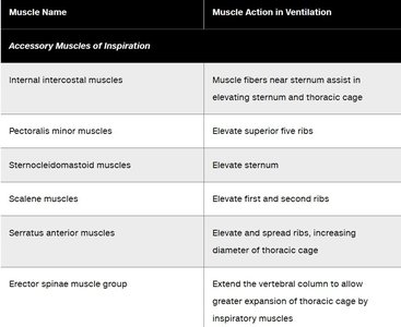
Accessory Muscles of Inspiration
Muscle Name | Muscle Action in Ventilation |
|---|---|
Internal intercostal muscles | Muscle fibers near sternum assist in elevating sternum and thoracic cage |
Pectoralis minor muscles | Elevate superior five ribs |
Sternocleidomastoid muscles | Elevate sternum |
Scalene muscles | Elevate first and second ribs |
Serratus anterior muscles | Elevate and spread ribs, increasing diameter of thoracic cage |
Erector spinae muscle group | Extend the vertebral column to allow greater expansion of thoracic cage by inspiratory muscles |
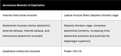
Accessory Muscles of Expiration
Muscle Name | Muscle Action in Ventilation |
|---|---|
Internal intercostal muscles | Lateral muscle fibers depress thoracic cage |
Abdominal muscles (rectus abdominis, external oblique, internal oblique, transversus abdominis) | Depress thoracic cage; compress abdominal contents, increasing intra-abdominal pressure and pushing the diaphragm superiorly |
Quadratus lumborum muscles | Fixate 12th rib |
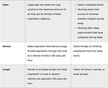
Nonrespiratory Movements
Types and Functions
Nonrespiratory movements are not directly involved in breathing but help maintain airway patency and alveolar inflation.
Movement | Definition | Function |
|---|---|---|
Yawn | Large sigh that takes the lung volume to the maximum amount of air that can be forcibly inhaled (inspiratory capacity) | Opens collapsed alveoli; minimizes alveolar collapse during sleep |
Sneeze | Deep inspiration followed by a large, forceful expiration through the nose at a velocity of about 100 miles per hour | Clears foreign or irritating substances from the nasal cavity |
Cough | Similar to a sneeze except the initial inspiration is small or absent; velocity can approach 500 miles per hour | Clears the larynx, trachea, or lower airways |
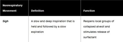
Nonrespiratory Movement | Definition | Function |
|---|---|---|
Sigh | A slow and deep inspiration that is held and followed by a slow expiration | Reopens local groups of collapsed alveoli and stimulates release of surfactant |
Physical Factors Affecting Pulmonary Ventilation
Airway Resistance
Anything that impedes airflow through the respiratory tract increases resistance.
Resistance decreases during inspiration (airways expand) and increases during expiration (airways narrow).
Bronchodilation (sympathetic stimulation) decreases resistance; bronchoconstriction (parasympathetic stimulation or irritants) increases resistance.
Alveolar Surface Tension
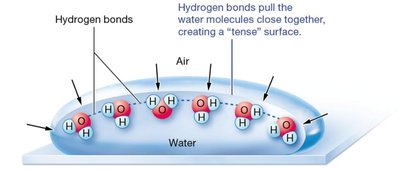
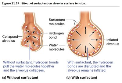
Water molecules in the alveoli are attracted to each other by hydrogen bonds, creating surface tension that tends to collapse the alveoli. Surfactant, produced by type II alveolar cells, disrupts these bonds and keeps alveoli open.
Pulmonary Compliance
Pulmonary compliance is the ability of the lungs and chest wall to stretch. It is determined by:
Degree of alveolar surface tension
Distensibility of elastic tissue in the lungs
Ability of the chest wall to move
Diseases that decrease surfactant, destroy elastic tissue, or cause fibrosis reduce compliance and make breathing more difficult.
Infant Respiratory Distress Syndrome (RDS)
Premature infants may lack sufficient surfactant, leading to alveolar collapse and difficulty breathing.
Treatment includes administration of surfactant and use of continuous positive airway pressure (CPAP).
Pulmonary Volumes and Capacities
Pulmonary Volumes
Tidal Volume (TV): Amount of air inspired or expired during normal quiet breathing (~500 mL).
Inspiratory Reserve Volume (IRV): Volume of air forcibly inspired after normal inspiration (2100–3300 mL).
Expiratory Reserve Volume (ERV): Volume of air forcibly expired after normal expiration (700–1200 mL).
Residual Volume (RV): Air remaining in lungs after maximal expiration, prevents lung collapse.
Pulmonary Capacities
Pulmonary Capacity | Average Value (Female, Male) | Description |
|---|---|---|
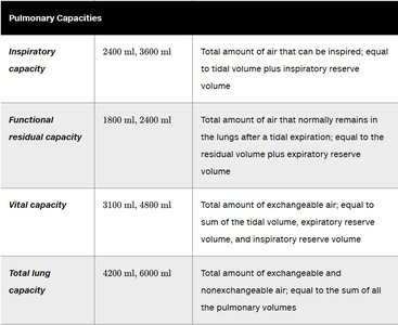
Inspiratory capacity | 2400 ml, 3600 ml | Total amount of air that can be inspired; equal to tidal volume plus inspiratory reserve volume |
Functional residual capacity | 1800 ml, 2400 ml | Total amount of air that normally remains in the lungs after a tidal expiration; equal to the residual volume plus expiratory reserve volume |
Vital capacity | 3100 ml, 4800 ml | Total amount of exchangeable air; equal to sum of the tidal volume, expiratory reserve volume, and inspiratory reserve volume |
Total lung capacity | 4200 ml, 6000 ml | Total amount of exchangeable and nonexchangeable air; equal to the sum of all the pulmonary volumes |
Summary of Pulmonary Ventilation Cycle
The process of pulmonary ventilation is a continuous cycle driven by pressure gradients created by changes in thoracic volume. This ensures efficient gas exchange and maintenance of homeostasis.
Additional info: Spirometry is used to measure pulmonary volumes and capacities, which are important for diagnosing respiratory diseases. Anatomical dead space refers to the portion of air that does not participate in gas exchange because it remains in the conducting airways.
