 Back
BackReflex Arcs and Their Classification: Study Notes for Anatomy & Physiology
Study Guide - Smart Notes
 Tailored notes based on your materials, expanded with key definitions, examples, and context.
Reflex Arcs: Introduction and Overview
Definition and Basic Concepts
A reflex is a rapid, automatic response to a specific stimulus, essential for maintaining homeostasis and protecting the body from harm. Reflexes are mediated by neural pathways known as reflex arcs. The interneuron is a neuron that transmits impulses between sensory and motor neurons, often serving as the integration center in the reflex arc.
Reflex Arc: The neuronal pathway that controls a reflex action, typically consisting of five main steps.
Example: Touching a hot object and immediately withdrawing your hand is a classic reflex action.
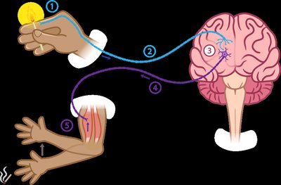
Five Steps of a Reflex Arc
Receptor: Detects the stimulus (e.g., pain, temperature, pressure).
Sensory Neuron: Transmits the afferent impulse from the receptor to the central nervous system (CNS).
Integration Center: Consists of one or more interneurons in the CNS that process the information and direct the response.
Motor Neuron: Conducts efferent impulses from the integration center to an effector.
Effector: Muscle or gland that responds to the efferent impulse by contracting or secreting.
Example Question: Which of the following is the first step in a reflex arc? Answer: Receptor
Classification of Reflex Arcs
Criteria for Classification
Reflexes can be classified based on their development, response type, and complexity.
Development:
Innate Reflex: Genetically programmed during natural development (e.g., knee-jerk reflex).
Acquired Reflex: Learned, complex motor patterns (e.g., conditioned taste aversion).
Response Type:
Somatic Reflex: Involuntary control of skeletal muscles (e.g., withdrawal from pain).
Autonomic (Visceral) Reflex: Involuntary control of smooth muscle, cardiac muscle, or glands (e.g., pupillary light reflex).
Complexity:
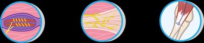
Monosynaptic Reflex: Involves a single synapse between a sensory and a motor neuron (e.g., stretch reflex).
Polysynaptic Reflex: Involves multiple synapses with one or more interneurons (e.g., withdrawal reflex).
Criterion | Type | Example |
|---|---|---|
Development | Innate | Knee-jerk reflex |
Development | Acquired | Conditioned taste aversion |
Response Type | Somatic | Withdrawal from pain |
Response Type | Autonomic | Pupillary light reflex |
Complexity | Monosynaptic | Stretch reflex |
Complexity | Polysynaptic | Flexor reflex |

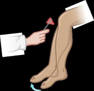
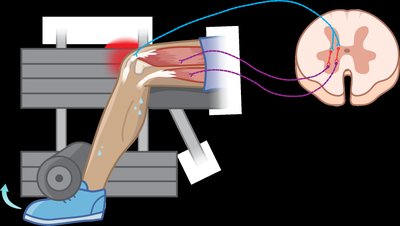
Stretch Reflex and Reciprocal Inhibition
Stretch Reflex
The stretch reflex is a monosynaptic reflex that helps prevent muscle strain and tear injuries. It is initiated by muscle spindles in response to muscle stretching, causing the muscle to contract.
Purpose: Prevents overstretching and maintains muscle tone.
Mechanism: Muscle spindles detect stretch, sensory neurons transmit the signal to the spinal cord, and motor neurons cause the muscle to contract.
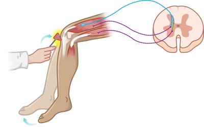
Reciprocal Inhibition
During the stretch reflex, neurons controlling the antagonistic muscle are inhibited, allowing smooth movement and preventing opposition to the contraction.
Purpose: Ensures coordinated movement by relaxing antagonistic muscles.
Mechanism: Involves polysynaptic pathways for inhibition of antagonists.
Example: The knee-jerk reflex contracts the quadriceps while inhibiting the hamstrings, helping maintain balance.
Tendon Reflex and Reciprocal Activation
Tendon Reflex
The tendon reflex is a polysynaptic reflex that prevents tendon injury by causing muscle relaxation in response to excessive tension detected by Golgi tendon organs.
Purpose: Protects tendons and muscles from damage due to excessive force.
Mechanism: Golgi tendon organs sense tension, leading to inhibition of the contracting muscle and activation of the antagonist.
Reciprocal Activation
During the tendon reflex, antagonistic muscles are activated to relieve tension on the tendon, ensuring joint stability and preventing injury.
Example: During heavy lifting, the tendon reflex relaxes the quadriceps and activates the hamstrings to prevent tendon rupture.
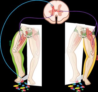
Flexor and Crossed-Extensor Reflexes
Flexor (Withdrawal) Reflex
The flexor reflex is a rapid, polysynaptic withdrawal of a body part from a painful stimulus. It is an ipsilateral reflex, meaning the response occurs on the same side as the stimulus.
Purpose: Protects the body from injury by rapidly withdrawing from harmful stimuli.
Mechanism: Flexor muscles contract to move the limb away from the source of pain.
Crossed-Extensor Reflex
The crossed-extensor reflex occurs simultaneously with the flexor reflex, causing extension of the opposite limb to maintain balance. It is a contralateral reflex, with the response occurring on the side opposite to the stimulus.
Purpose: Maintains posture and balance during withdrawal movements.
Mechanism: While one limb withdraws, the other extends to support body weight.
Summary Table: Types of Reflexes
Reflex Type | Stimulus | Response | Synapses | Example |
|---|---|---|---|---|
Stretch Reflex | Muscle stretch | Muscle contraction | Monosynaptic | Knee-jerk reflex |
Tendon Reflex | Excessive tension | Muscle relaxation | Polysynaptic | Golgi tendon reflex |
Flexor Reflex | Painful stimulus | Withdrawal | Polysynaptic | Hand withdrawal from heat |
Crossed-Extensor Reflex | Painful stimulus | Extension of opposite limb | Polysynaptic | Stepping on a sharp object |
Additional info: Reflexes are essential for survival, allowing the body to respond rapidly to potentially harmful stimuli without the need for conscious thought. They are also used clinically to assess the integrity of the nervous system.
