 Back
BackRenal System: Structure, Function, and Regulation
Study Guide - Smart Notes
 Tailored notes based on your materials, expanded with key definitions, examples, and context.
Renal System Overview
Importance of the Kidneys
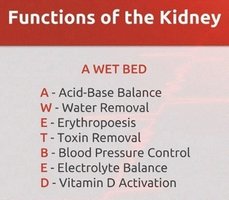
The kidneys are vital organs responsible for maintaining homeostasis by regulating the composition and volume of blood. They perform several essential functions:
Salt and water balance: Controls fluid volume and osmolarity.
Regulation of blood ion concentrations: Maintains electrolyte balance.
Acid-base balance: Adjusts pH by excreting hydrogen ions and reabsorbing bicarbonate.
Excretion of metabolic wastes, drugs, and toxins: Removes harmful substances from the body.
Production of erythropoietin (EPO): Stimulates red blood cell production.
Production of renin: Regulates blood pressure.
Activation of vitamin D: Converts vitamin D to its active form for calcium regulation.
Anatomy of the Urinary System
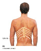
Gross Anatomy of the Kidneys
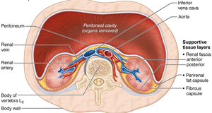
The kidneys are bean-shaped, retroperitoneal organs located in the superior lumbar region, spanning from the 12th thoracic vertebra to the 3rd lumbar vertebra. The right kidney is slightly lower due to the liver. Each kidney weighs about 150 g and measures 12 x 6 x 3 cm. Adrenal glands sit atop each kidney.
Supportive Tissue Layers
Three layers of supportive tissue surround each kidney:
Renal capsule: Fibrous layer adhering directly to the kidney surface, providing a strong barrier.
Perirenal fat capsule: Cushions and helps hold the kidney in place.
Renal fascia: Dense connective tissue that surrounds the kidney and adrenal gland, anchoring them.
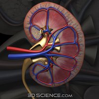
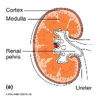
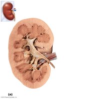
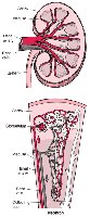
Internal Anatomy of the Kidney
The kidney's internal structure consists of three main regions:
Cortex: The outer region where filtration occurs.
Medulla: Contains medullary or renal pyramids, which appear striped due to parallel bundles of tubules and blood vessels. Separated by renal columns.
Renal pelvis: Funnel-shaped tube continuous with the ureter. Major and minor calyces collect urine from the pyramids.
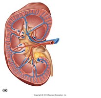
Blood and Nerve Supply
The kidneys receive about 1/4 of the total systemic cardiac output (~1.2 L/min). Renal arteries branch to reach the cortex, and venous branches drain blood back via the same route. The renal plexus, primarily composed of sympathetic fibers, regulates renal blood flow by adjusting the diameter of renal arterioles.
Nephron Structure and Types
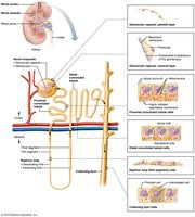
Nephron Anatomy
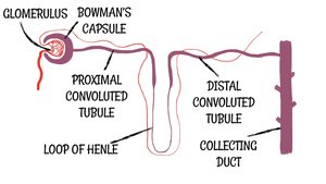
Each kidney contains approximately one million nephrons, the functional units responsible for urine formation. The nephron consists of:
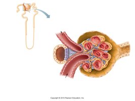
Renal corpuscle: Includes the glomerulus and Bowman's capsule.
Proximal convoluted tubule (PCT): Reabsorbs water, ions, and nutrients.
Nephron loop (Loop of Henle): Establishes concentration gradients.
Distal convoluted tubule (DCT): Further adjusts ion and water balance.
Collecting duct: Final site for water and ion regulation.
Nephron Types
Cortical nephrons: Make up 85% of nephrons; located mostly in the cortex; responsible for most filtration.
Juxtamedullary nephrons: Make up 15%; have long loops extending deep into the medulla; crucial for concentrating urine.
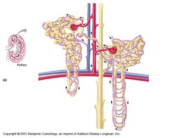
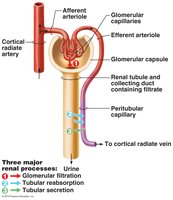
Microcirculation of the Nephron
The glomerulus is specialized for filtration and is both fed and drained by arterioles. The afferent arteriole has a larger diameter, creating high pressure for filtration. Peritubular capillaries and vasa recta arise from efferent arterioles and are involved in reabsorption and maintaining medullary concentration gradients.
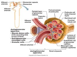
Juxtaglomerular Complex (JGC)
The JGC is located at the junction of the early distal convoluted tubule and afferent/efferent arterioles. It regulates renal function and blood pressure via:
Granular (JG) cells: Mechanoreceptors that secrete renin.
Macula densa cells: Chemoreceptors/osmoreceptors that monitor filtrate and adjust glomerular filtration rate (GFR).
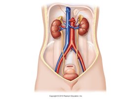
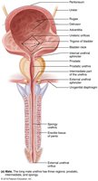
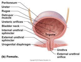
Urinary Bladder and Micturition Pathways
Male and Female Urinary Systems
The urinary bladder stores urine and is capable of holding up to 1 liter. The trigone is a triangular area important clinically due to its susceptibility to infection. The internal urethral sphincter is formed by smooth muscle, while the external urethral sphincter is formed by skeletal muscle.
Renal Physiology: Filtration, Reabsorption, and Secretion
Nephron as the Functional Unit
The nephron filters several liters of fluid from the bloodstream daily, removing toxins, metabolic wastes, and excess ions while returning needed materials to the blood.
Major Renal Processes
Glomerular filtration: Passive, nonselective process driven by hydrostatic pressure.
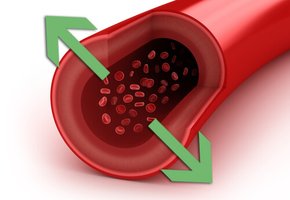
Tubular reabsorption: Selective process returning nutrients, ions, and water to the blood.
Tubular secretion: Active process removing additional wastes and excess ions.
Filtrate vs. Urine
Filtrate: Plasma minus proteins; formed in the glomerulus.
Urine: Filtrate minus nutrients, essential ions, and most water; final waste product.
Glomerular Filtration
Glomerular filtration is highly efficient due to the permeability of the filtration membrane and high glomerular blood pressure (55 mm Hg). The filtration membrane consists of three layers:
Fenestrated capillary endothelium
Basement membrane
Visceral membrane of glomerular capsule (podocytes)
Filtration Membrane Function
Molecules < 3 nm (water, glucose, amino acids, nitrogenous wastes) pass easily.
Molecules 3-5 nm pass with difficulty.
Molecules > 5 nm are usually not filtered.
Basement membrane proteins restrict passage of most plasma proteins.
Retention of plasma proteins maintains colloid osmotic pressure.
Presence of proteins or RBCs in urine suggests membrane damage.

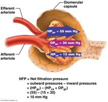
Net Filtration Pressure (NFP)
NFP is the pressure responsible for filtrate formation and is calculated as:
HPg: Glomerular hydrostatic pressure (pushes out of blood)
OPg: Glomerular osmotic pressure (pulls back into blood)
HPc: Capsular hydrostatic pressure (pushes back into blood)
Formula:
Example:
Glomerular Filtration Rate (GFR)
GFR is the total amount of filtrate formed per minute by both kidneys (~125 ml/min). It depends on:
Total surface area for filtration
Filtration membrane permeability
Net filtration pressure
GFR is directly proportional to NFP. A decrease in glomerular blood pressure of only ~15% can stop filtration.

Regulation of Glomerular Filtration Rate
Regulatory Influences
Three main mechanisms regulate GFR:
Renal autoregulation (intrinsic): Kidneys adjust their own blood flow and nephron function.
Neural controls (extrinsic): Sympathetic nervous system adjusts blood flow during stress.
Renin-angiotensin system (extrinsic): Hormonal regulation of blood pressure and GFR.
Renal Autoregulation
Renal autoregulation maintains a constant GFR with systemic blood pressure between 80-180 mm Hg. Below 80 mm Hg, extrinsic regulation takes over.
Myogenic Mechanism
Vascular smooth muscle contracts when stretched, regulating afferent arteriole diameter in response to blood pressure changes.

Tubuloglomerular Feedback Mechanism
Macula densa cells monitor NaCl content in filtrate. High osmolarity triggers vasoconstriction; low osmolarity allows vasodilation.

Summary Table: Kidney Functions
Function | Description |
|---|---|
Acid-Base Balance | Regulates blood pH by excreting H+ and reabsorbing HCO3- |
Water Removal | Controls fluid volume and osmolarity |
Erythropoiesis | Stimulates RBC production via EPO |
Toxin Removal | Excretes metabolic wastes and drugs |
Blood Pressure Control | Regulates via renin-angiotensin system |
Electrolyte Balance | Maintains ion concentrations |
Vitamin D Activation | Converts vitamin D to active form |
Key Equations
Net Filtration Pressure:
Glomerular Filtration Rate: GFR is directly proportional to NFP
Additional info:
Juxtamedullary nephrons are more abundant in animals adapted to arid environments (e.g., camels) to maximize water conservation.
Presence of proteins or RBCs in urine is a clinical indicator of kidney pathology.
