 Back
Respiratory Physiology: Mechanics and Gas Exchange
Study Guide - Smart Notes
 Tailored notes based on your materials, expanded with key definitions, examples, and context.
Mechanics of Breathing
Pulmonary Ventilation
Pulmonary ventilation is the process of moving air into and out of the lungs, consisting of two phases: inspiration (inhalation) and expiration (exhalation). This process is driven by pressure changes within the thoracic cavity, which are caused by changes in lung volume.
Inspiration: Gases flow into the lungs as thoracic volume increases and pressure decreases.
Expiration: Gases exit the lungs as thoracic volume decreases and pressure increases.
Pressure Relationships in the Thoracic Cavity
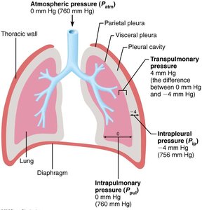
Several pressures are important in pulmonary ventilation:
Atmospheric Pressure (Patm): The pressure exerted by air surrounding the body, typically 760 mm Hg at sea level.
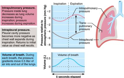
Intrapulmonary Pressure (Ppul): The pressure within the alveoli, fluctuates with breathing but always equalizes with Patm.
Intrapleural Pressure (Pip): The pressure within the pleural cavity, always negative relative to Patm and Ppul (usually about 4 mm Hg less than Ppul).
Transpulmonary Pressure: The difference between Ppul and Pip; it keeps the lungs inflated.
Pneumothorax and Atelectasis
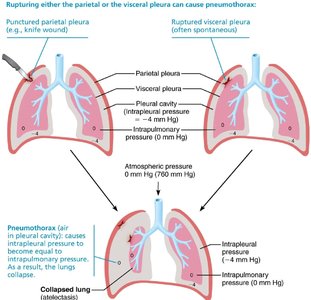
Pneumothorax is the presence of air in the pleural cavity, which can result from trauma or spontaneous rupture of the pleura. This condition causes Pip to equalize with Patm, leading to lung collapse (atelectasis).
Treatment: Removal of air with chest tubes allows the lung to reinflate as the pleura heals.
Physical Principles of Pulmonary Ventilation
Boyle’s Law
Boyle’s law describes the inverse relationship between the pressure and volume of a gas at constant temperature:
As volume increases, pressure decreases, and vice versa.
Mathematically:
Sequence of Events During Inspiration and Expiration
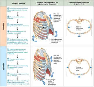
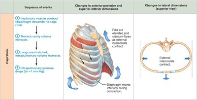
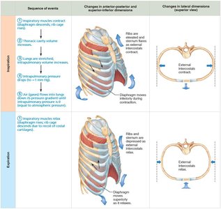
Inspiration is an active process involving contraction of the diaphragm and external intercostal muscles, increasing thoracic volume and decreasing intrapulmonary pressure. Expiration is usually passive, resulting from relaxation of inspiratory muscles and elastic recoil of the lungs.
Detailed Steps of Inspiration
Inspiratory muscles contract (diaphragm descends; rib cage rises).
Thoracic cavity volume increases.
Lungs are stretched; intrapulmonary volume increases.
Intrapulmonary pressure drops (to -1 mm Hg).
Air flows into lungs down its pressure gradient until Ppul = Patm.
Detailed Steps of Expiration
Inspiratory muscles relax (diaphragm rises; rib cage descends).
Thoracic cavity volume decreases.
Elastic lungs recoil passively; intrapulmonary volume decreases.
Intrapulmonary pressure rises (to +1 mm Hg).
Air flows out of lungs down its pressure gradient until Ppul = Patm.
Changes in Intrapulmonary and Intrapleural Pressures
During inspiration, both intrapulmonary and intrapleural pressures decrease, facilitating air entry. During expiration, these pressures increase, driving air out of the lungs. The pressure gradients move approximately 0.5 liters of air in and out with each breath.
Physical Factors Influencing Pulmonary Ventilation
Airway Resistance
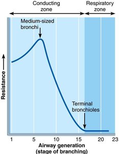
Airway resistance is primarily due to friction in the airways. It is usually insignificant due to large airway diameters and extensive branching, but can increase in conditions like asthma.
Relationship: , where F is flow, ΔP is pressure gradient, and R is resistance.
Alveolar Surface Tension
Surface tension in the alveoli is reduced by surfactant, a lipid-protein complex produced by type II alveolar cells. Surfactant prevents alveolar collapse by decreasing the attraction between water molecules.
Insufficient surfactant in premature infants leads to Infant Respiratory Distress Syndrome (IRDS).
Lung Compliance
Lung compliance is a measure of the lung's ability to stretch and expand. High compliance means the lungs can expand easily, while low compliance requires more energy for breathing.
Mathematically:
Decreased by fibrosis, reduced surfactant, or decreased thoracic flexibility.
Assessing Ventilation
Respiratory Volumes and Capacities
Several volumes and capacities are measured to assess lung function:
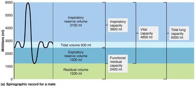
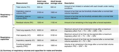
Tidal Volume (TV): Air moved in/out with each breath (~500 ml).
Inspiratory Reserve Volume (IRV): Air that can be forcibly inhaled beyond TV (2100–3200 ml).
Expiratory Reserve Volume (ERV): Air that can be forcibly exhaled beyond TV (1000–1200 ml).
Residual Volume (RV): Air remaining in lungs after forced expiration (1200 ml).
Measurement | Adult Male Avg. Value | Adult Female Avg. Value | Description |
|---|---|---|---|
Total Volume (TV) | 500 ml | 500 ml | Amount of air inhaled/exhaled with each breath under resting conditions |
Inspiratory Reserve Volume (IRV) | 3100 ml | 1900 ml | Amount of air that can be forcibly inhaled after normal tidal volume inspiration |
Expiratory Reserve Volume (ERV) | 1200 ml | 700 ml | Amount of air that can be forcibly exhaled after normal tidal volume expiration |
Residual Volume (RV) | 1200 ml | 1100 ml | Amount of air remaining in the lungs after forced expiration |
Total Lung Capacity (TLC) | 6000 ml | 4200 ml | Maximum amount of air contained in lungs after maximum inspiratory effort |
Vital Capacity (VC) | 4800 ml | 3100 ml | Maximum amount of air that can be expired after a maximum inspiratory effort |
Inspiratory Capacity (IC) | 3600 ml | 2400 ml | Maximum amount of air that can be inspired after a normal tidal expiration |
Functional Residual Capacity (FRC) | 2400 ml | 1800 ml | Volume of air remaining in the lungs after a normal tidal expiration |
Dead Space
Anatomical dead space is the volume of air that does not participate in gas exchange (~150 ml). Alveolar dead space is due to nonfunctional alveoli. Total dead space is the sum of anatomical and alveolar dead space.
Pulmonary Function Tests
Obstructive diseases: Increased airway resistance (e.g., bronchitis); TLC, FRC, RV may increase.
Restrictive diseases: Reduced TLC due to disease or fibrosis; VC, TLC, FRC, RV decline.
Forced Vital Capacity (FVC): Gas forcibly expelled after deep breath.
Forced Expiratory Volume (FEV): Gas expelled during specific time intervals of FVC.
Alveolar Ventilation
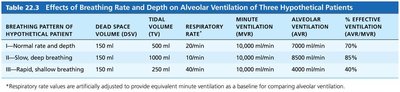
Minute ventilation is the total amount of gas entering or leaving the respiratory tract per minute. Alveolar ventilation rate (AVR) is a better indicator of effective ventilation, calculated as:
Increasing tidal volume is more effective than increasing frequency for improving AVR.
Gas Exchange
Basic Properties of Gases
Dalton’s Law: The total pressure of a gas mixture is the sum of the partial pressures of each gas.
Henry’s Law: The amount of gas dissolved in a liquid is proportional to its partial pressure and solubility.
Partial Pressures of Atmospheric and Alveolar Gases
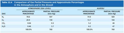
Gas | Atmosphere % | Atmosphere (mm Hg) | Alveoli % | Alveoli (mm Hg) |
|---|---|---|---|---|
N2 | 78.6 | 597 | 74.9 | 569 |
O2 | 20.9 | 159 | 13.7 | 104 |
CO2 | 0.04 | 0.3 | 5.2 | 40 |
H2O | 0.46 | 3.7 | 6.2 | 47 |
External Respiration
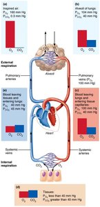
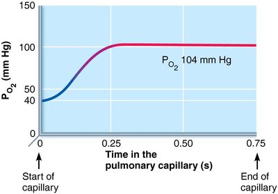
External respiration is the exchange of O2 and CO2 between alveoli and blood. It is influenced by partial pressure gradients, membrane thickness, surface area, and ventilation-perfusion coupling.
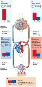
O2 diffuses from alveoli (PO2 = 104 mm Hg) to blood (PO2 = 40 mm Hg).
CO2 diffuses from blood (PCO2 = 45 mm Hg) to alveoli (PCO2 = 40 mm Hg).
Factors Affecting External Respiration
Membrane Thickness: Increased thickness (e.g., edema) impairs gas exchange.
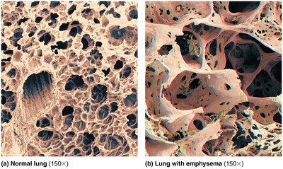
Surface Area: Reduced in diseases like emphysema, tumors, or mucus blockage.
Ventilation-Perfusion Coupling
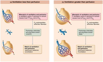
Optimal gas exchange requires matching of alveolar ventilation and pulmonary blood perfusion. Local autoregulatory mechanisms adjust arteriolar and bronchiolar diameters in response to O2 and CO2 levels.
Internal Respiration
Internal respiration is the exchange of gases between systemic blood and tissues. The gradients are reversed compared to external respiration:
O2 moves from blood (PO2 = 100 mm Hg) to tissues (PO2 = 40 mm Hg).
CO2 moves from tissues (PCO2 = 45 mm Hg) to blood (PCO2 = 40 mm Hg).
Additional info: These notes cover the core physiological mechanisms of breathing and gas exchange, including clinical correlations relevant to pulmonary pathology and assessment.
