 Back
BackRespiratory System: Anatomy and Physiology Study Guide
Study Guide - Smart Notes
 Tailored notes based on your materials, expanded with key definitions, examples, and context.
Respiratory Anatomy
Structure of the Respiratory System
The respiratory system is divided into the upper and lower respiratory tracts, each with distinct anatomical structures and functions essential for breathing and gas exchange.
Upper respiratory tract: Includes the nasal cavity, pharynx, and larynx.
Lower respiratory tract: Comprises the trachea, its branches, and the lungs.
Upper Respiratory System Structures
The upper respiratory tract conducts air and, in some regions, food and fluids. The pharynx is subdivided into three regions:
Nasopharynx: Air passage from posterior nares to soft palate.
Oropharynx: Passage for air and food from soft palate to superior margin of epiglottis.
Laryngopharynx: Passage for air and food from superior margin of epiglottis to cricoid cartilage.
Larynx (Voice Box)
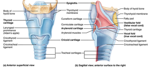
The larynx is located inferior to the laryngopharynx and is responsible for sound production and airway protection. It consists of three paired and three single cartilages, mostly composed of hyaline cartilage except for the epiglottis.
Key cartilages: Thyroid, cricoid, arytenoid, corniculate, cuneiform, and epiglottis.
Function: Protects airway during swallowing and produces vocal sounds.
Lower Respiratory System Structures
Lungs
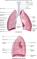
The lungs are paired, soft, spongy organs responsible for gas exchange. Each lung has an apex, base, and lobes (2 on the left, 3 on the right).
Lobes: Right lung (superior, middle, inferior), Left lung (superior, inferior).
Function: Houses bronchi, bronchioles, and alveoli for gas exchange.
Trachea
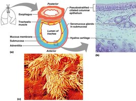
The trachea, or windpipe, is lined with ciliated mucus-secreting cells and supported by C-shaped rings of hyaline cartilage. It divides into primary bronchi, which further branch into secondary, tertiary bronchi, and bronchioles, ending in alveolar sacs.
Epithelium: Pseudostratified ciliated columnar epithelium.
Function: Conducts air to the lungs and filters particles.
Alveoli
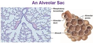
Alveoli are balloon-like protrusions with simple squamous epithelium, forming the blood/air barrier and serving as the primary site for gas exchange.
Structure: Alveolar sacs, alveolar ducts, and pores.
Function: Facilitates oxygen and carbon dioxide exchange between air and blood.
Respiratory Physiology
Mechanics of Breathing
Pulmonary ventilation consists of inspiration and expiration, driven by changes in thoracic cavity volume and pressure.
Inspiration: Diaphragm contracts and flattens, thoracic cavity expands, air enters lungs.
Expiration: Muscles relax, lungs recoil, air is expelled.
Boyle’s Law
Boyle’s Law describes the relationship between gas pressure and volume at constant temperature:
Formula: (where P is pressure, V is volume, and k is a constant)
Implication: As lung volume increases, pressure decreases, allowing air to flow in; as volume decreases, pressure increases, forcing air out.
Respiratory Volumes
Respiratory volumes measure the amount of air moved during breathing:
Tidal Volume (TV): ~500 mL per breath at rest.
Inspiratory Reserve Volume (IRV): Air forcibly inhaled after normal inhalation.
Expiratory Reserve Volume (ERV): Air forcibly exhaled after normal exhalation.
Vital Capacity (VC):
Pneumothorax
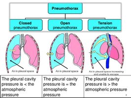
Pneumothorax occurs when air enters the pleural cavity, causing lung collapse and increased pressure. Types include closed (spontaneous), open, and tension pneumothorax.
Closed: Air leaks into pleural space, chest wall and pleura intact.
Open: "Sucking chest wound", chest wall and pleura not intact.
Tension: Air enters pleural space but cannot escape, pressure builds, may cause mediastinal shift and tracheal deviation.
Treatment: Needle decompression to aspirate air.
Summary Table: Respiratory Volumes
Volume | Description |
|---|---|
Tidal Volume (TV) | Amount of air inhaled/exhaled per breath at rest (~500 mL) |
Inspiratory Reserve Volume (IRV) | Air forcibly inhaled after normal inhalation |
Expiratory Reserve Volume (ERV) | Air forcibly exhaled after normal exhalation |
Vital Capacity (VC) | Maximum air exhaled after maximal inspiration (TV + IRV + ERV) |
Key Terms
Pharynx: Passage for air, food, and fluids.
Larynx: Voice box, airway protection.
Trachea: Windpipe, conducts air to lungs.
Alveoli: Site of gas exchange.
Pneumothorax: Air in pleural cavity, lung collapse.
