 Back
BackRespiratory System: Gas Exchange, Volumes, and Capacities 23-2
Study Guide - Smart Notes
 Tailored notes based on your materials, expanded with key definitions, examples, and context.
Respiratory Volumes and Capacities
Overview of Lung Volumes and Capacities
Respiratory volumes and capacities are essential measurements for assessing ventilation and diagnosing pulmonary disorders. They describe the amount of air the lungs can hold and the changes in lung volume during different phases of breathing.
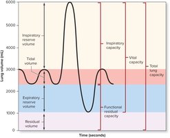
Tidal Volume (TV): The amount of air taken into or expelled out of the lungs during a quiet breath.
Inspiratory Reserve Volume (IRV): The amount of air taken into the lungs during a forced inspiration, following a quiet inspiration. IRV is a measure of lung compliance.
Expiratory Reserve Volume (ERV): The amount of air expelled from the lungs during a forced expiration, following a quiet expiration. ERV is a measure of lung and chest wall elasticity.
Residual Volume (RV): The amount of air left in the lungs following a forced expiration.
Capacities Derived from Volumes
Lung capacities are combinations of two or more lung volumes and represent the total ability to inspire or expire air.
Inspiratory Capacity: TV + IRV; total ability to inspire.
Functional Residual Capacity: ERV + RV; amount of air typically left in lungs after quiet expiration.
Vital Capacity: TV + IRV + ERV; maximum amount of air that can be forcefully expired after a forced inspiration.
Total Lung Capacity: TV + IRV + ERV + RV; maximum amount of air the lungs can hold.
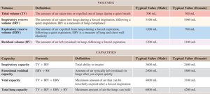
Volume | Definition | Typical Value (Male) | Typical Value (Female) |
|---|---|---|---|
Tidal volume (TV) | Amount of air taken in or expelled during quiet breath | 500 mL | 500 mL |
Inspiratory reserve volume (IRV) | Amount of air taken in during forced inspiration after quiet inspiration | 3100 mL | 1900 mL |
Expiratory reserve volume (ERV) | Amount of air expelled during forced expiration after quiet expiration | 1200 mL | 700 mL |
Residual volume (RV) | Amount of air left in lungs after forced expiration | 1200 mL | 1100 mL |
Capacity | Formula | Definition | Typical Value (Male) | Typical Value (Female) |
|---|---|---|---|---|
Inspiratory capacity | TV + IRV | Total ability to inspire | 3600 mL | 2400 mL |
Functional residual capacity | ERV + RV | Amount of air typically left in lungs after quiet expiration | 2400 mL | 1800 mL |
Vital capacity | TV + IRV + ERV | Maximum amount of air that can be forcefully expired after forced inspiration | 4800 mL | 3100 mL |
Total lung capacity | TV + IRV + ERV + RV | Maximum amount of air the lungs can hold | 6000 mL | 4200 mL |
Four Processes of Respiration
Overview of Respiration
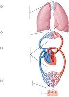
Respiration consists of four integrated processes that ensure the exchange and transport of gases necessary for cellular function.
Pulmonary Ventilation: Movement of air into (inspiration) and out of (expiration) the lungs.
External Respiration: Diffusion of gases between alveoli and blood.
Transport of Respiratory Gases: Movement of O2 and CO2 in the blood between lungs and tissues.
Internal Respiration: Diffusion of gases between blood and tissue cells.
Chemical Principles of Gas Exchange
Partial Pressure and Dalton’s Law
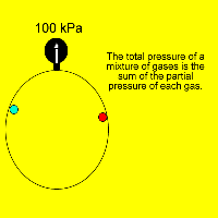
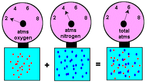
Gas exchange is governed by the partial pressures of gases and Dalton’s law. Partial pressure is the pressure exerted by each gas in a mixture, and each gas moves independently down its partial pressure gradient.
Dalton’s Law: The total pressure of a mixture of gases is equal to the sum of the partial pressures of each gas.
Formula:
Example: Nitrogen is 78.6% of air; at 760 mm Hg, its partial pressure is mm Hg.
Gas Exchange in the Lungs and Tissues
External Respiration (Pulmonary Gas Exchange)
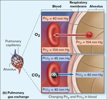
Pulmonary gas exchange is the movement of O2 and CO2 between alveolar air and pulmonary capillary blood, driven by partial pressure gradients.
Oxygen: PO2 in alveoli = 104 mm Hg; PO2 in blood entering capillaries = 40 mm Hg. Oxygen diffuses from alveoli to blood.
Carbon Dioxide: PCO2 in alveoli = 40 mm Hg; PCO2 in blood = 45 mm Hg. CO2 diffuses from blood to alveoli.
Internal Respiration (Tissue Gas Exchange)
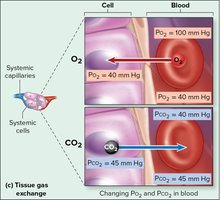
Tissue gas exchange is the movement of O2 and CO2 between systemic capillary blood and tissue cells.
Oxygen: PO2 in systemic capillaries = 95 mm Hg; PO2 in cells = 40 mm Hg. Oxygen diffuses from blood to cells.
Carbon Dioxide: PCO2 in cells = 45 mm Hg; PCO2 in capillaries = 40 mm Hg. CO2 diffuses from cells to blood.
Comparison of Gas Partial Pressures in Atmosphere and Alveoli
Atmospheric vs. Alveolar Gas Composition
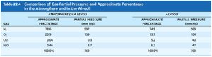
The composition of gases in the alveoli differs from atmospheric air due to gas exchange, humidification, and mixing with air in the dead space.
Gas | Approximate Percentage (Atmosphere) | Partial Pressure (Atmosphere, mm Hg) | Approximate Percentage (Alveoli) | Partial Pressure (Alveoli, mm Hg) |
|---|---|---|---|---|
N2 | 78.6% | 597 | 74.9% | 569 |
O2 | 20.9% | 159 | 13.7% | 104 |
CO2 | 0.04% | 0.3 | 5.2% | 40 |
H2O | 0.46% | 3.7 | 6.2% | 47 |
Total | 100% | 760 | 100% | 760 |
Factors Influencing Gas Exchange
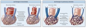
Ventilation-Perfusion Coupling
Ventilation-perfusion coupling is the matching of alveolar ventilation with pulmonary blood perfusion. It ensures optimal gas exchange by adjusting bronchiolar and arteriolar diameters in response to local PO2 and PCO2 levels.
Perfusion: Blood flow reaching alveoli.
Ventilation: Amount of gas reaching alveoli.
Regulation: PO2 controls perfusion by changing arteriolar diameter; PCO2 controls ventilation by changing bronchiolar diameter.
Example: Increased PCO2 causes bronchioles to dilate; decreased PO2 causes arterioles to constrict.
 Additional info: Ventilation-perfusion mismatch can lead to impaired gas exchange and hypoxemia.
