 Back
BackSensation and Sensory Receptors: Structure, Function, and Pathways
Study Guide - Smart Notes
 Tailored notes based on your materials, expanded with key definitions, examples, and context.
Topic 8: Sensation
Introduction to Sensation and Perception
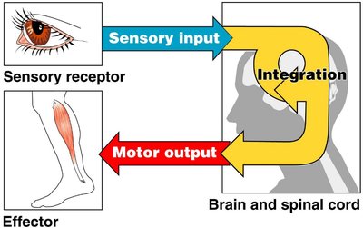
Sensation is the process by which sensory receptors detect and transmit information about the environment to the central nervous system (CNS). Perception is the conscious awareness and interpretation of these sensory signals. Sensory receptors are specialized cells or cell processes that monitor internal and external conditions, providing the CNS with essential information for survival and homeostasis.
Sensory receptors: Specialized cells that detect specific stimuli (e.g., light, sound, temperature, pressure).
Sensation: The raw information carried by a sensory pathway.
Perception: The conscious awareness and interpretation of a sensation.
Receptive field: The area monitored by a single receptor cell; larger fields reduce localization accuracy.
Sensory Pathways and Transduction
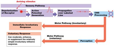
All sensory pathways begin with the depolarization of sensory receptors. Physical or chemical stimuli are detected and converted into electrical signals through a process called sensory transduction. The strength of the stimulus determines the strength of the receptor potential, which can lead to the generation of action potentials that travel to the CNS.
Sensory transduction: Conversion of a stimulus into an electrical signal (receptor potential).
Receptor potential: A graded change in membrane potential; stronger stimuli produce stronger potentials.
Action potentials: All-or-nothing electrical signals triggered if the receptor potential reaches threshold.
Example: Sensory Transduction in Taste
The process of sensory transduction can be illustrated by the sense of taste. When a sugar molecule binds to a taste receptor cell, it triggers a signal transduction pathway, leading to changes in ion channel activity and membrane potential. This results in neurotransmitter release and the generation of action potentials in sensory neurons, which the brain interprets as taste intensity.
Step 1: Stimulus (e.g., sugar) binds to receptor on taste bud.
Step 2: Signal transduction pathway is activated.
Step 3: Ion channels open/close, altering membrane potential (receptor potential).
Step 4: Neurotransmitter release increases action potential rate in sensory neuron.

Receptor Potentials vs. Action Potentials
Receptor potentials are graded and vary in intensity with the strength of the stimulus, unlike action potentials, which are all-or-nothing events. This allows sensory systems to encode stimulus intensity before the signal is transmitted to the CNS.
Receptor potential: Graded, variable in intensity, occurs in receptor cell.
Action potential: All-or-nothing, occurs in sensory neuron if threshold is reached.
Sensory Adaptation
Sensory adaptation is the reduction in sensitivity to a constant stimulus. It can occur at the level of the receptor (peripheral adaptation) or within the CNS (central adaptation).
Peripheral adaptation: Receptor response declines with constant stimulation (e.g., temperature adaptation).
Central adaptation: CNS inhibition reduces awareness of a stimulus even if sensory neurons are still active (e.g., adapting to a persistent smell).
Types of Human Senses
General vs. Special Senses
The human body has two main categories of senses: general senses and special senses. General senses are distributed throughout the body and include touch, temperature, pain, pressure, vibration, and proprioception. Special senses are localized to specific organs and include vision, hearing, balance, taste, and smell.
General senses: Detected by receptors in skin and tissues; lack specialized organs.
Special senses: Detected by specialized organs (e.g., eyes, ears, tongue, nose).
Classification of Sensory Receptors
Overview of Sensory Receptor Types
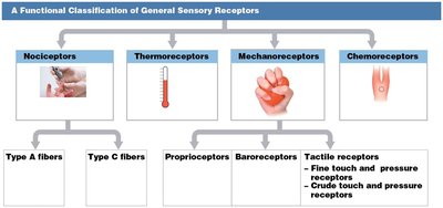
Sensory receptors are classified by the type of stimulus they detect. The four main types are nociceptors, thermoreceptors, mechanoreceptors, and chemoreceptors.
Receptor Type | Stimulus Detected | Subtypes |
|---|---|---|
Nociceptors | Pain | Type A fibers (fast), Type C fibers (slow) |
Thermoreceptors | Temperature | Cold and warm receptors |
Mechanoreceptors | Physical distortion | Proprioceptors, Baroreceptors, Tactile receptors |
Chemoreceptors | Chemical concentration | Monitor pH, CO2, O2 |
Nociceptors (Pain Receptors)
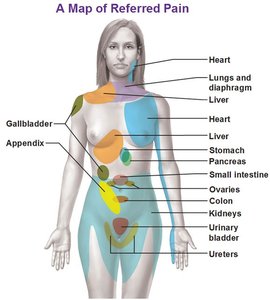
Nociceptors are free nerve endings that detect pain. They have large receptive fields and broad sensitivity, and are found in skin, joints, bones, and internal organs. Pain can be referred, meaning it is perceived in a different location from its source. Nociceptors do not adapt quickly, ensuring that pain is noticed and addressed.
Type A fibers: Myelinated, fast pain (sharp, localized).
Type C fibers: Unmyelinated, slow pain (dull, aching).
Referred pain: Pain from internal organs perceived at a distant site.
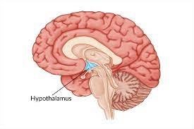
Thermoreceptors (Temperature Receptors)
Thermoreceptors are free nerve endings that detect changes in temperature. They are found in the dermis, skeletal muscles, liver, and hypothalamus. Cold receptors are more numerous than warm receptors, but both types lack structural differences.
Monitor environmental and internal temperature changes.
Play a role in thermoregulation and homeostasis.
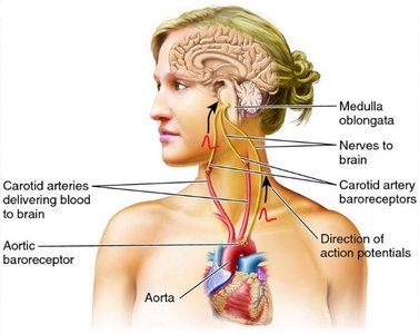
Chemoreceptors
Chemoreceptors detect changes in the concentration of specific chemicals in body fluids. They are essential for reflexive control of respiration and cardiovascular function, monitoring pH, carbon dioxide, and oxygen levels in the blood and cerebrospinal fluid.
Located in the medulla oblongata, carotid bodies, and aortic bodies.
Trigger reflexive changes in breathing and cardiovascular activity.
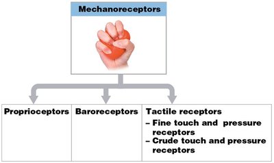
Mechanoreceptors
Mechanoreceptors respond to physical distortion of their plasma membranes, such as stretching, compression, or twisting. They have mechanically gated ion channels and are divided into three main types: proprioceptors, baroreceptors, and tactile receptors.
Proprioceptors: Monitor position of joints and muscles.
Baroreceptors: Detect pressure changes in blood vessels and organs.
Tactile receptors: Detect touch, pressure, and vibration.

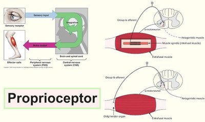
Proprioceptors
Proprioceptors are the most complex general sensory receptors, providing information about the position and movement of the body. They are found in muscles, tendons, and joints, and are essential for coordination and balance.
Examples: Muscle spindles, Golgi tendon organs.
Baroreceptors
Baroreceptors detect pressure changes in blood vessels and hollow organs. They are free nerve endings that respond to stretching or recoiling of elastic tissues, altering the rate of action potential generation. Baroreceptors play a key role in regulating blood pressure, lung expansion, and organ volume.
Locations: Carotid sinus, aortic sinus, lungs, colon, digestive tract, urinary bladder.
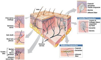
Tactile Receptors
Tactile receptors provide sensations of touch, pressure, and vibration. They are abundant in the skin and include several types, each with distinct structures and functions.
Free nerve endings: Simplest, respond to multiple stimuli, most common in skin.
Root hair plexus: Detect hair movement, adapt rapidly.
Tactile discs (Merkel discs): Fine touch and pressure, very sensitive, small receptive fields.
Tactile corpuscles (Meissner's corpuscles): Fine touch, pressure, low-frequency vibration, adapt rapidly, found in sensitive areas.
Lamellar corpuscles (Pacinian corpuscles): Deep pressure, high-frequency vibration, fast-adapting, large receptors.
Bulbous corpuscles (Ruffini corpuscles): Pressure and distortion of deep dermis, little adaptation.
Structure of Tactile Receptors
Receptor | Location | Stimulus | Adaptation |
|---|---|---|---|
Free nerve endings | Skin, mucous membranes | Touch, pain, temperature | Variable |
Root hair plexus | Hair follicles | Hair movement | Rapid |
Tactile discs | Stratum basale | Fine touch, pressure | Slow |
Tactile corpuscles | Dermal papillae | Fine touch, vibration | Rapid |
Lamellar corpuscles | Deep dermis, organs | Deep pressure, vibration | Rapid |
Bulbous corpuscles | Deep dermis | Pressure, skin stretch | Slow |
Summary Table: Sensory Receptor Types and Functions
Receptor Type | Stimulus | Location | Adaptation |
|---|---|---|---|
Nociceptors | Pain | Skin, joints, organs | Slow |
Thermoreceptors | Temperature | Dermis, muscles, hypothalamus | Fast |
Chemoreceptors | Chemicals (pH, CO2, O2) | Blood vessels, brain | Variable |
Mechanoreceptors | Physical distortion | Skin, muscles, organs | Variable |
Key Concepts for Review
Differentiate between general and special senses.
Describe the structure and function of each type of sensory receptor and their subtypes (nociceptors, thermoreceptors, mechanoreceptors, chemoreceptors).
Explain the process of sensory transduction and the difference between receptor potentials and action potentials.
Understand sensory adaptation and its physiological significance.
