 Back
BackSensation, Perception, and Sensory Integration in Anatomy & Physiology
Study Guide - Smart Notes
 Tailored notes based on your materials, expanded with key definitions, examples, and context.
Sensation and Perception
Definitions and Processes
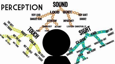
Sensation and perception are fundamental concepts in anatomy and physiology, describing how the body detects and interprets stimuli from the environment. Sensation refers to the awareness of a stimulus, requiring four processes: stimulation, transduction, conduction, and translation. Perception is the conscious awareness and interpretation of those sensations.
Sensation: Involves detection of stimuli and conversion to neural signals.
Perception: Involves interpretation of sensory information in the brain.
Processes:
Stimulation: Activation of sensory receptors.
Transduction: Conversion of stimulus to electrical signal.
Conduction: Transmission of signal to CNS.
Translation: Interpretation in the brain.
Example: Touching a hot object activates thermoreceptors, which send signals to the brain, resulting in the perception of heat.
Organization of the Nervous System
CNS and PNS Structure
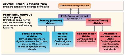
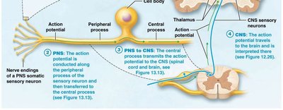
The nervous system is divided into the Central Nervous System (CNS) and Peripheral Nervous System (PNS). The CNS integrates information, while the PNS links the CNS to the rest of the body and performs motor and sensory functions.
CNS: Brain and spinal cord.
PNS: Cranial and spinal nerves.
Divisions of PNS:
Sensory (afferent) division: Carries sensory signals to CNS.
Motor (efferent) division: Carries motor signals from CNS.
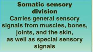
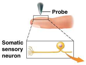
Somatic Sensory Division: Carries general sensory signals from muscles, bones, joints, and skin, as well as special sensory signals.
Sensory Transduction
Conversion of Stimulus to Electrical Signal
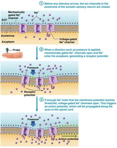
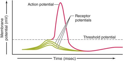
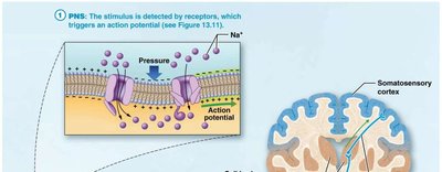
Sensory transduction is the process by which a physical stimulus is converted into an electrical signal in the nervous system. This occurs at the sensory receptor level and involves the generation of receptor potentials and, if threshold is reached, action potentials.
Receptor Potentials: Produced when a stimulus is detected, often due to influx of Na+ ions.
Action Potentials: Generated when receptor potential reaches threshold, allowing the signal to be conducted to the CNS.
Adaptation: When a stimulus persists, action potentials may stop. There are two types: rapid and slow adaptation.
Classification of Sensory Receptors
Types and Functions
Sensory receptors are classified based on microscopic structure, location, and the type of stimulus detected.
Microscopic Structure: Free nerve endings, encapsulated endings, or separate cells.
Location:
Exteroreceptors: Detect external stimuli.
Interoreceptors: Detect internal stimuli.
Type of Stimulus Detected: Mechanoreceptors (touch, pressure), thermoreceptors (temperature), nociceptors (pain), photoreceptors (light), chemoreceptors (chemicals).
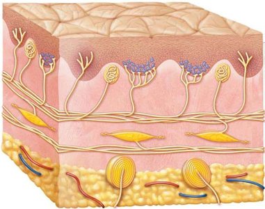
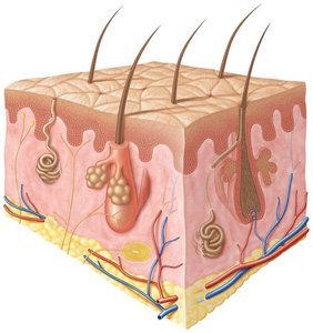
Mechanoreceptors in Skin
Mechanoreceptors are specialized for detecting mechanical changes such as touch, pressure, and vibration.
Merkel cell fibers: Slow adapting; fine touch.
Meissner (tactile) corpuscles: Rapid adaptation; fine touch.
Ruffini endings: Slow-adapting; stretch and movement.
Pacinian (lamellated) corpuscles: Rapid adaptation; vibration and pressure.
Hair follicle receptors: Free nerve endings at base of follicle.
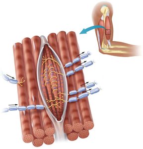
Proprioceptors: Sense movement and position of joints (kinesthetic sense); found in muscle spindles, tendon organs, and joints.
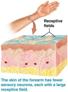
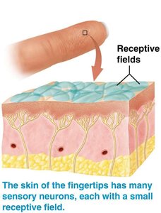
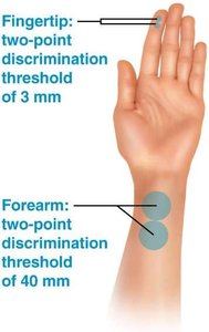
Sensory Neurons: Speed and Receptive Fields
Conduction Speed and Touch Discrimination
Sensory neurons vary in conduction speed and receptive field size, affecting touch discrimination.
Fast Conduction: Large axon diameter and myelination; associated with proprioception.
Slow Conduction: Small axon diameter and less myelination; associated with pain and temperature.
Touch Discrimination: Ability to identify type and source of touch stimulus increases with higher density of small-field receptors.
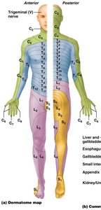
Dermatomes and Pain Perception
Mapping Sensory Pathways and Pain Types
Dermatomes are regions of skin innervated by specific spinal nerves, useful for mapping sensory pathways. Nociceptors provide information about tissue damage and disease symptoms. Pain can be localized as somatic (superficial or deep), visceral, or referred, and can be fast or slow.
Somatic Pain: Originates from skin, muscles, or joints.
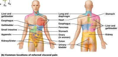
Visceral Pain: Originates from internal organs; often referred to other body regions.
Referred Pain: Pain perceived at a location other than the site of origin.
Detection and Perception of Sensation
Neural Pathways and CNS Integration
Sensory information is carried to the CNS by (pseudo)unipolar neurons. Visceral sensory information terminates in the brainstem/diencephalon, while somatic sensory information terminates in the somatosensory cortex of the cerebrum.
Somatic Sensory Neurons: Carry information from skin, muscles, and joints.
Somatosensory Cortex: Responsible for conscious perception of somatic sensations.
CNS Integration and Homeostasis
Role of Hypothalamus and Autonomic Centers
The CNS integrates sensory information in the hypothalamus and autonomic centers, regulating functions such as thirst, hunger, heat, respiratory, cardiac, digestive, thermoregulation, micturition, and water balance.
Hypothalamus: Integrates sensory input and regulates homeostatic functions.
Autonomic Centers: Control involuntary functions such as heart rate, respiration, digestion, and thermoregulation.
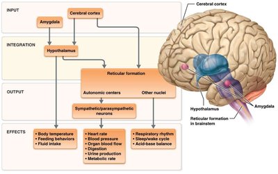
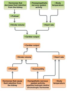
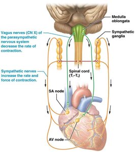
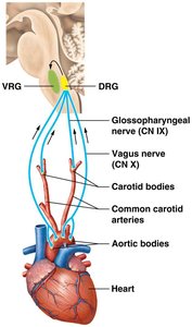
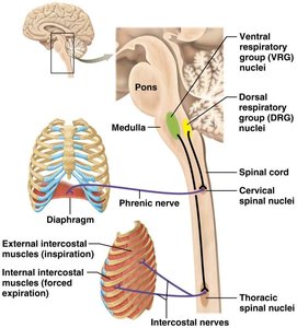
Cardiac and Respiratory Function
Neural Control Centers
Heart rate and force of contraction are controlled by centers in the brainstem. The dorsal and ventral respiratory group nuclei regulate the rate and depth of ventilation.
Cardiac Function: Controlled by medulla oblongata and sympathetic/parasympathetic nerves.
Respiratory Function: Controlled by pons and medulla, affecting inspiration and expiration.
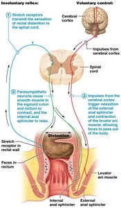
Digestive Function and Defecation Reflex
Enteric Nervous System and Voluntary Control
The enteric nervous system controls most digestive processes. Defecation is regulated by parasympathetic function and requires the primary motor cortex for voluntary control.
Defecation Reflex: Involuntary and voluntary pathways coordinate the expulsion of feces.
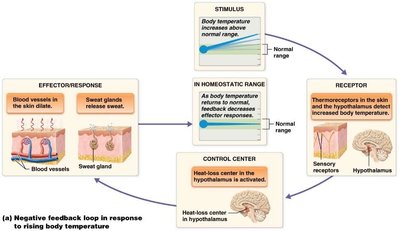
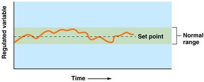
Thermoregulation
Heat-Loss and Heat-Gain Centers
Thermoregulation is managed by centers in the hypothalamus, which control responses to maintain body temperature within a normal range.
Heat-Loss Center: Activates mechanisms to reduce body temperature.
Heat-Gain Center: Activates mechanisms to increase body temperature.
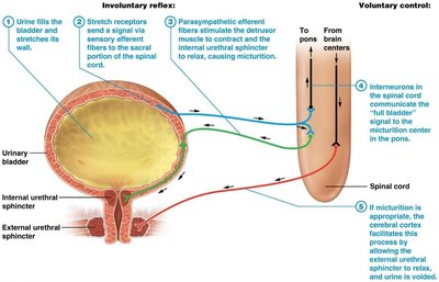
Micturition (Urination)
Neural Control of Bladder Function
Micturition centers are located in the pons of the brainstem and are controlled via parasympathetic function. Voluntary control requires the primary motor cortex.
Involuntary Reflex: Stretch receptors in the bladder trigger parasympathetic responses.
Voluntary Control: Cerebral cortex facilitates relaxation of sphincters for urination.
Water Balance
Osmoreceptors and Baroreceptors
Water balance is regulated by osmoreceptors and baroreceptors, which send information to thirst centers in the hypothalamus. Fluid intake is stimulated by various physiological cues.
Osmoreceptors: Detect changes in osmolarity.
Baroreceptors: Detect changes in blood pressure.
Nervous and Endocrine System Integration
Hypothalamus and Pituitary Relationship
The hypothalamus and pituitary gland have a close anatomical relationship, allowing for efficient communication and control between the nervous and endocrine systems.
Hypothalamus: Regulates endocrine functions via the pituitary.
Integration: Enables coordinated responses to maintain homeostasis.
Summary Table: Mechanoreceptors in Skin
The following table summarizes the main mechanoreceptors found in the skin and their functions:
Mechanoreceptor | Function |
|---|---|
Merkel cell fiber | Discriminative touch with fine spatial resolution |
Tactile corpuscle | Discriminative touch with less spatial resolution |
Ruffini ending (bulbous corpuscle) | Stretch and movement |
Lamellated corpuscle | Vibration and deep pressure |
