 Back
BackSensorimotor System: Sensory Receptors, Pathways, and Reflexes
Study Guide - Smart Notes
 Tailored notes based on your materials, expanded with key definitions, examples, and context.
Sensorimotor System Overview
Introduction to the Sensorimotor System
The sensorimotor system integrates sensory input and motor output, allowing the body to respond to internal and external stimuli. It includes both voluntary (somatic) and involuntary (autonomic) divisions, coordinating functions such as movement, posture, and visceral regulation.
Somatic Division: Controls voluntary movements and transmits sensory information from skin, muscles, and joints to the brain.
Autonomic Division: Regulates involuntary functions, including heart rate, smooth muscle activity, and glandular secretion. It is further divided into sympathetic and parasympathetic systems.
Sensory Receptors and Sensation
Classification of Sensory Receptors
Sensory receptors are specialized cells or nerve endings that detect changes in the environment (stimuli) and initiate nerve impulses. Sensation is the awareness of these stimuli, while perception is their interpretation in the brain.
By Stimulus Type:
Mechanoreceptors: Respond to touch, pressure, vibration, and stretch.
Thermoreceptors: Detect temperature changes.
Photoreceptors: Respond to light (e.g., in the retina).
Chemoreceptors: Detect chemicals (e.g., taste, smell, blood chemistry).
Nociceptors: Respond to pain-causing stimuli (e.g., extreme temperature, pressure, chemicals).
By Location:
Exteroceptors: Detect external stimuli (touch, pressure, pain, temperature); found in skin and special sense organs.
Interoceptors (Visceroceptors): Detect internal stimuli (chemical changes, stretch, temperature) in viscera and blood vessels.
Proprioceptors: Detect stretch and position in muscles, tendons, joints, and connective tissue; inform the brain about body position and movement.
By Structural Complexity:
Simple Receptors: General senses (touch, pain, temperature, muscle sense); found throughout the body.
Complex Receptors: Special senses (vision, hearing, equilibrium, smell, taste); located in specialized organs.
Structural Types of Sensory Receptors
Nonencapsulated (Free) Nerve Endings: Abundant in epithelia and connective tissues; respond to temperature, pain, and light touch.
Encapsulated Nerve Endings: Enclosed in connective tissue capsules; respond to pressure, vibration, and stretch.
Examples of Sensory Receptors
Receptor Type | Location | Stimulus Type |
|---|---|---|
Free nerve endings | Most body tissues, especially connective tissue and epithelia | Temperature, pain, pressure |
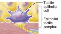
Merkel (tactile) discs | Basal layer of epidermis | Light pressure |
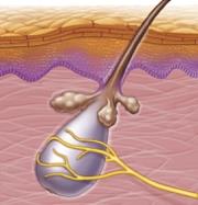
Hair follicle receptors | Surrounding hair follicles | Hair deflection |
Meissner's (tactile) corpuscles | Dermal papillae of hairless skin | Light pressure, discriminative touch |
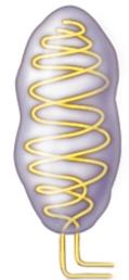
Pacinian (lamellar) corpuscles | Dermis, hypodermis, periostea, etc. | Deep pressure, vibration |
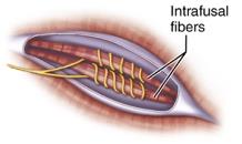
Muscle spindles | Skeletal muscles | Muscle stretch, length |
Tendon organs | Tendons | Tendon stretch, tension |
Neural Integration in Sensory Systems
Three Levels of Neural Integration
Sensory information is processed at three main levels:
Receptor Level: Sensory receptors detect stimuli and generate graded potentials.
Circuit Level: Processing occurs in ascending pathways to the brain.
Perceptual Level: Sensory information is interpreted in cortical sensory centers.
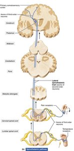
Ascending Sensory Pathways
Major Ascending Pathways
Ascending sensory pathways transmit information from receptors to the brain for conscious perception or unconscious processing.
Dorsal Column–Medial Lemniscal Pathway: Fine touch, vibration, proprioception; synapses in the medulla and thalamus before reaching the somatosensory cortex.
Spinothalamic Pathway: Pain, temperature, crude touch, pressure; synapses in the spinal cord and thalamus before reaching the cortex.
Spinocerebellar Pathway: Muscle and tendon stretch; information sent to the cerebellum for coordination (not conscious sensation).
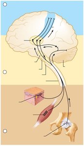
Descending Motor Pathways
Direct and Indirect Pathways
Descending motor pathways deliver efferent impulses from the brain to the spinal cord, controlling voluntary and involuntary movements.
Direct (Pyramidal) Pathways: Originate from pyramidal cells in the primary motor cortex; regulate fast and fine (skilled) movements via the lateral and ventral corticospinal tracts.
Indirect (Extrapyramidal) Pathways: Include all other motor pathways; regulate balance, posture, coarse limb movements, and head/neck/eye movements.
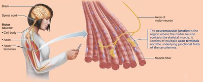
Motor Endings and Skeletal Muscle Contraction
Neuromuscular Junction and Muscle Contraction
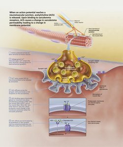
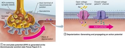
The neuromuscular junction is the site where a motor neuron communicates with a skeletal muscle fiber, initiating muscle contraction.
Events at the Neuromuscular Junction:
Acetylcholine (ACh) is released from the neuron and binds to receptors on the muscle cell membrane (sarcolemma).
This causes depolarization and triggers an action potential in the muscle fiber.
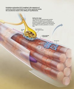
Excitation-Contraction Coupling: The action potential travels along the sarcolemma and T-tubules, leading to Ca2+ release from the sarcoplasmic reticulum and muscle contraction.
Cross Bridge Cycle: Myosin heads bind to actin, pull thin filaments toward the center of the sarcomere, and generate force for contraction.

Reflex Activity
Reflex Arc and Types of Reflexes
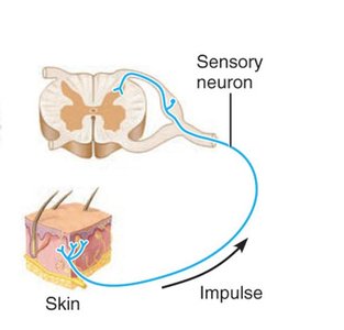
A reflex is a rapid, involuntary response to a stimulus. Reflexes are mediated by neural pathways called reflex arcs, which consist of five basic components:
Receptor
Sensory neuron
Integration center
Motor neuron
Effector
Somatic Reflexes: Activate skeletal muscle.
Autonomic (Visceral) Reflexes: Activate smooth or cardiac muscle or glands.
Stretch and Tendon Reflexes
These reflexes help maintain muscle tone and prevent muscle damage.
Stretch Reflex: Initiated by muscle spindle stretch; causes contraction of the stretched muscle and inhibition of its antagonist. Example: knee-jerk reflex.
Tendon Reflex: Initiated by tension in tendon organs; causes muscle relaxation and prevents excessive force.
Flexor and Crossed-Extensor Reflexes
Flexor (Withdrawal) Reflex: Initiated by a painful stimulus; causes withdrawal of the affected body part.
Crossed-Extensor Reflex: Accompanies the flexor reflex in weight-bearing limbs; maintains balance by extending the opposite limb.
Superficial Reflexes
Plantar Reflex: Tests integrity of spinal cord from L4 to S2; abnormal response (Babinski's sign) indicates corticospinal tract damage.
Abdominal Reflex: Tests integrity of cord from T8 to T12; absence may indicate corticospinal tract lesions.
Additional info: These notes integrate content from chapters on the nervous system, sensory receptors, motor pathways, and muscle physiology, as outlined in the ANP college course syllabus.
