 Back
Sensory Pathways and the Somatic Nervous System: Structure, Function, and Clinical Relevance
Study Guide - Smart Notes
 Tailored notes based on your materials, expanded with key definitions, examples, and context.
Introduction to Sensory Pathways and the Somatic Nervous System
This chapter explores the organization and function of sensory and motor pathways within the nervous system, focusing on the general senses and the somatic nervous system (SNS). It distinguishes between the afferent (sensory) and efferent (motor) divisions, and introduces clinical relevance through conditions such as cerebral palsy.
Sensory and Motor Pathways
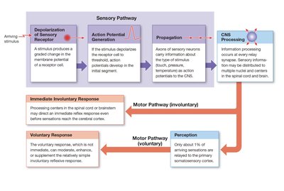
Overview of Sensory and Motor Pathways
Sensory pathways relay information from sensory receptors to the central nervous system (CNS), while motor pathways transmit commands from the CNS to peripheral effectors. The somatic nervous system controls voluntary skeletal muscle contractions, whereas the autonomic nervous system (ANS) regulates involuntary actions of smooth muscle, cardiac muscle, glands, and adipose tissue.
Sensory Pathways: Involve a sequence of neurons transmitting signals from receptors to the CNS.
Motor Pathways: Carry commands from the CNS to effectors, either voluntarily (skeletal muscle) or involuntarily (visceral effectors).
Components of the Afferent and Efferent Divisions
Afferent Division
The afferent division consists of somatic and visceral sensory pathways. Somatic sensory information is sent to the primary somatosensory cortex or cerebellum, while visceral sensory information is directed to reflex centers in the brainstem and diencephalon.
Somatic Sensory Pathways: Convey information about the external environment and body position.
Visceral Sensory Pathways: Monitor internal organ function.
Efferent Division
The efferent division includes the somatic motor pathways (voluntary control of skeletal muscles) and the autonomic motor pathways (involuntary control of visceral effectors).
Sensory Receptors and Their Function
Types and Roles of Sensory Receptors
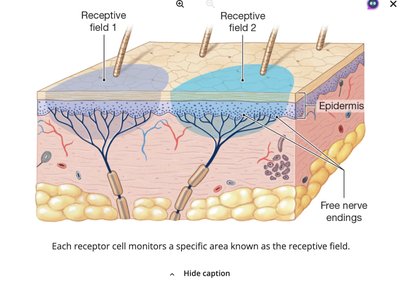
Sensory receptors are specialized cells or cell processes that detect specific stimuli and convert them into electrical signals (action potentials) through a process called transduction. The area monitored by a single receptor is its receptive field.
Sensation: The arrival of sensory information at the CNS.
Perception: Conscious awareness of a sensation.
Receptor Specificity: Each receptor responds to a particular type of stimulus.
Transduction and Sensory Coding
Transduction is the conversion of a stimulus into an action potential. The frequency and pattern of action potentials encode the strength, duration, and type of stimulus, a process known as sensory coding.
Generator Potential: A depolarizing receptor potential that can trigger action potentials in sensory neurons.
Labeled Line: The pathway from a receptor to a specific region of the cortex, determining the perceived modality.
Tonic and Phasic Receptors
Receptors can be classified by their response patterns:
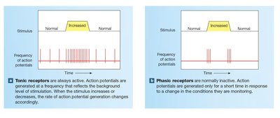
Tonic Receptors: Always active; frequency of action potentials reflects stimulus intensity.
Phasic Receptors: Normally inactive; respond with bursts of activity to changes in stimulus conditions.
Adaptation in Sensory Receptors
Adaptation is a reduction in receptor sensitivity during constant stimulation. It can be peripheral (at the receptor) or central (within the CNS). Tonic receptors adapt slowly, while phasic receptors adapt quickly.
Classification of General Sensory Receptors
By Location
Exteroceptors: Monitor external environment.
Proprioceptors: Monitor body position and movement.
Interoceptors: Monitor internal organ function.
By Stimulus Type
Nociceptors: Detect pain from harmful stimuli.
Thermoreceptors: Detect temperature changes.
Mechanoreceptors: Detect physical distortion (touch, pressure, vibration).
Chemoreceptors: Detect chemical composition of body fluids.
Mechanoreceptors: Structure and Function
Tactile Receptors
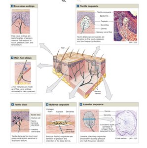
Tactile receptors provide sensations of touch, pressure, and vibration. They range from simple free nerve endings to complex structures with accessory cells.
Receptor Type | Location/Structure | Function |
|---|---|---|
Free nerve endings | Between epidermal cells | Touch, pressure, pain, temperature |
Root hair plexus | Base of hair follicles | Detect hair movement |
Tactile discs | Stratum basale of skin | Fine touch, shape, texture |
Bulbous corpuscles | Deep dermis | Pressure, skin distortion |
Lamellar corpuscles | Dermis, fasciae, joint capsules | Deep pressure, vibration |
Tactile corpuscles | Eyelids, lips, fingertips, nipples, genitalia | Fine touch, low-frequency vibration |
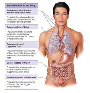
Baroreceptors
Baroreceptors detect pressure changes in the walls of blood vessels and hollow organs. They play a critical role in regulating cardiovascular and respiratory functions, as well as reflexes in the digestive and urinary systems.
Proprioceptors
Proprioceptors monitor the position of joints, tension in tendons and ligaments, and the state of muscle contraction. They are essential for body position awareness and coordination.
Muscle Spindles: Monitor muscle length and trigger stretch reflexes.
Golgi Tendon Organs: Monitor tension in tendons during muscle contraction.
Joint Capsule Receptors: Detect pressure, tension, and movement at joints.
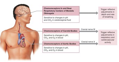
Chemoreceptors
Chemoreceptors respond to changes in the concentration of specific chemicals in body fluids. They are important for the reflexive control of respiration and cardiovascular function, monitoring pH, CO2, and O2 levels in blood and cerebrospinal fluid.
Clinical Application: Cerebral Palsy
Overview
Cerebral palsy (CP) is a congenital neurological condition resulting from brain injury before, during, or shortly after birth. It is characterized by impaired motor function, delayed developmental milestones, and often affects speech and movement. The specific symptoms depend on the regions of the brain affected.
Etiology: Brain injury due to hypoxia, trauma, or infection.
Symptoms: Motor impairment, spasticity, difficulty with voluntary movement, and sometimes cognitive or sensory deficits.
Clinical Relevance: Understanding sensory and motor pathways is essential for diagnosing and managing CP and similar neurological conditions.
Summary Table: General Sensory Receptors
Receptor Type | Stimulus Detected | Location | Adaptation |
|---|---|---|---|
Nociceptors | Pain (harmful stimuli) | Skin, joints, periosteum, blood vessels | Slow (tonic) |
Thermoreceptors | Temperature | Dermis, muscles, liver, hypothalamus | Fast (phasic) |
Mechanoreceptors | Touch, pressure, vibration, stretch | Skin, blood vessels, organs | Varies |
Chemoreceptors | Chemical composition | Blood vessels, brainstem | Peripheral and central |
Key Terms and Concepts
Transduction: Conversion of a stimulus into an action potential.
Receptive Field: Area monitored by a single receptor cell.
Adaptation: Decrease in sensitivity to a constant stimulus.
Tonic Receptor: Slow-adapting, always active receptor.
Phasic Receptor: Fast-adapting, normally inactive receptor.
Labeled Line: Pathway from receptor to cortex, determining modality.
