 Back
Sensory Pathways and the Somatic Nervous System: Structure and Function
Study Guide - Smart Notes
 Tailored notes based on your materials, expanded with key definitions, examples, and context.
Sensory Pathways and the Somatic Nervous System
Introduction
The sensory pathways and the somatic nervous system (SNS) are essential for detecting environmental changes and coordinating voluntary muscle movements. This chapter explores the structure, function, and classification of sensory receptors, the organization of sensory and motor pathways, and the integration of sensory input with motor output.
Sensory and Motor Pathways
Overview of Sensory and Motor Pathways
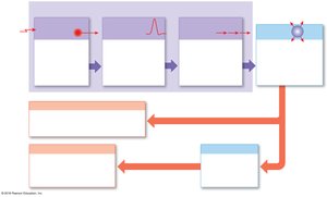
Sensory pathways relay information from receptors to the central nervous system (CNS), while motor pathways transmit commands from the CNS to peripheral effectors such as skeletal muscles. The afferent division carries sensory information, and the efferent division carries motor commands.
Sensory receptors: Specialized cells or processes that monitor specific conditions inside or outside the body.
Action potentials: Generated when receptors are stimulated, transmitting information along sensory pathways.
Processing: Occurs at every relay synapse, distributing sensory information to multiple CNS centers.
Immediate involuntary response: Reflexes may be triggered before conscious perception occurs.
Voluntary response: Initiated by the cerebral cortex, can enhance or override reflexes.
Sensory Receptors
Types and Functions of Sensory Receptors
Sensory receptors are either specialized neuron processes or cells monitored by sensory neurons. They detect stimuli and convert them into electrical signals (action potentials).
Sensation: The arrival of sensory information at the CNS.
Perception: Conscious awareness of a sensation.
General and Special Senses
General senses: Include temperature, pain, touch, pressure, vibration, and proprioception.
Special senses: Include olfaction (smell), gustation (taste), vision (sight), equilibrium (balance), and hearing. These are provided by specialized receptors in sense organs.
Detection and Interpretation of Stimuli
Receptor specificity: Each receptor is sensitive to a particular type of stimulus.
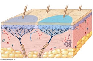
Receptive field: The area monitored by a single receptor cell. Larger fields make localization of stimuli more difficult.
Transduction: Conversion of a stimulus into an action potential.
Adaptation of Sensory Receptors
Adaptation: Reduction in sensitivity to a constant stimulus.
Peripheral adaptation: Occurs at the receptor level (PNS).
Central adaptation: Occurs in the CNS.
Tonic receptors: Always active, slow to adapt (e.g., pain receptors).
Phasic receptors: Normally inactive, fast-adapting, respond to changes in stimulus intensity.
General Sensory Receptors
Classification by Location and Stimulus
Exteroceptors: Monitor external environment.
Proprioceptors: Monitor positions of muscles and joints.
Interoceptors: Monitor internal organs.
Nociceptors: Detect pain from temperature extremes, mechanical damage, or chemicals.
Thermoreceptors: Detect temperature changes.
Mechanoreceptors: Detect physical distortion (stretch, compression, twisting).
Chemoreceptors: Detect chemical concentrations in body fluids.
Tactile Receptors in the Skin
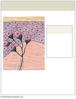
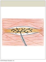
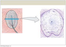
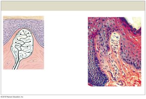
There are six main types of tactile receptors, each with distinct structure and function:
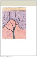
Free nerve endings: Sensitive to touch, pressure, pain, and temperature; found between epidermal cells.
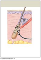
Root hair plexus: Detect hair movement; adapt rapidly.
Tactile discs (Merkel discs): Fine touch and pressure; sensitive to shape and texture; small receptive fields.
Bulbous corpuscles (Ruffini): Detect pressure and skin distortion; located in deep dermis; tonic receptors.
Lamellar corpuscles (Pacinian): Detect deep pressure and high-frequency vibration; fast-adapting.
Tactile corpuscles (Meissner): Detect fine touch, pressure, and low-frequency vibration; adapt quickly; abundant in sensitive areas.
Baroreceptors and Proprioceptors
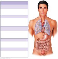
Baroreceptors: Monitor pressure changes in organs such as blood vessels, lungs, digestive tract, colon, and bladder.
Proprioceptors: Found in joints, tendons, ligaments, and muscles; monitor body position and movement.
Chemoreceptors
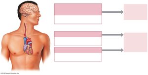
Chemoreceptors respond to water- and lipid-soluble substances dissolved in body fluids. They monitor pH, CO2, and O2 levels in arterial blood at carotid and aortic bodies, and in the medulla oblongata for cerebrospinal fluid.
Sensory Pathways
Organization of Sensory Pathways
First-order neuron: Delivers sensations to the CNS.
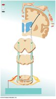
Second-order neuron: Interneuron in the spinal cord or brainstem; crosses to the opposite side (decussation).
Third-order neuron: Located in the thalamus; relays information to the cerebral cortex for awareness.
Major Somatic Sensory Pathways
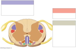
Spinothalamic pathway: Carries crude touch, pressure, pain, and temperature sensations.
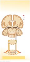
Posterior column pathway: Carries fine touch, vibration, pressure, and proprioception.
Spinocerebellar pathway: Conveys proprioceptive information to the cerebellum (not consciously perceived).
Visceral Sensory Pathways
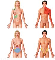
Visceral sensory information is collected by interoceptors in thoracic and abdominopelvic organs and relayed via cranial nerves V, VII, IX, and X to the solitary nucleus in the medulla oblongata.
Somatic Motor Pathways
Organization of Somatic Motor Pathways
Upper motor neuron: Cell body in CNS; synapses on lower motor neuron; can facilitate or inhibit movement.
Lower motor neuron: Cell body in brainstem or spinal cord; axon extends to skeletal muscle; triggers contraction.
Major Motor Pathways
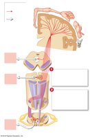
Corticospinal pathway (pyramidal system): Voluntary control of skeletal muscles; begins in the primary motor cortex.
Medial pathway: Controls gross movements of neck, trunk, and proximal limbs.
Lateral pathway: Controls muscle tone and precise movements of distal limbs.
Basal Nuclei and Cerebellum
Basal nuclei: Provide background patterns for voluntary movements; influence motor cortex and reticulospinal tracts.
Cerebellum: Monitors proprioceptive, visual, and vestibular input; fine-tunes movements through practice and feedback.
Summary Table: Types of Sensory Receptors
Receptor Type | Stimulus Detected | Location | Adaptation |
|---|---|---|---|
Nociceptors | Pain (mechanical, thermal, chemical) | Skin, joints, periosteum, blood vessels | Slow |
Thermoreceptors | Temperature | Dermis, skeletal muscle, liver, hypothalamus | Fast |
Mechanoreceptors | Physical distortion | Skin, muscles, organs | Varies |
Chemoreceptors | Chemical concentration | Blood vessels, brain, organs | Fast |
Additional info: This guide expands on the original notes with definitions, examples, and a summary table for clarity and exam preparation.
