 Back
BackSensory Physiology: Mechanisms and Pathways
Study Guide - Smart Notes
 Tailored notes based on your materials, expanded with key definitions, examples, and context.
Sensory Physiology
Introduction to Sensory Physiology
Sensory physiology explores how organisms detect and interpret information from their environment through specialized cells and neural pathways. This field is crucial for understanding clinical symptoms, such as numbness or tingling, and for localizing neurological damage.
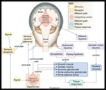
Sensory receptors detect specific types of stimuli and convert them into electrical signals.
These signals are transmitted via afferent pathways to the central nervous system (CNS), where they are processed and interpreted.
Clinical assessment of sensory symptoms helps localize damage within the nervous system.
Stimulus Strength: Intensity and Duration
Encoding Stimulus Properties
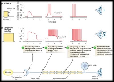
The nervous system encodes the intensity and duration of a stimulus through the number of receptors activated and the frequency of action potentials generated. The duration of a stimulus is reflected in the duration of action potential firing.
Tonic receptors: Slowly adapting; maintain firing as long as the stimulus is present (e.g., baroreceptors).
Phasic receptors: Rapidly adapting; fire at stimulus onset but cease if the stimulus remains constant (e.g., olfactory receptors).
Stimulus intensity is coded by the frequency of action potentials and the number of receptors activated.
Stimulus duration is coded by the duration of action potential firing.
Types of Sensory Information
Conscious and Subconscious Processing
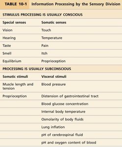
Sensory information can be processed consciously (e.g., touch, vision) or subconsciously (e.g., blood pressure, body fluid osmolarity). The table below summarizes the types of stimuli and their processing pathways.
Stimulus Processing | Special Senses | Somatic Senses |
|---|---|---|
Usually Conscious | Vision, Hearing, Taste, Smell, Equilibrium | Touch, Temperature, Pain, Itch, Proprioception |
Processing is Usually Subconscious | Somatic Stimuli | Visceral Stimuli |
Muscle length and tension, Proprioception | Blood pressure, Distension of GI tract, Blood glucose concentration, Internal body temperature, Osmolarity of body fluids, Lung inflation, pH of cerebrospinal fluid, pH and oxygen content of blood |
Somatosensory Pathways and Cortical Processing
Organization of Sensory Regions in the Brain
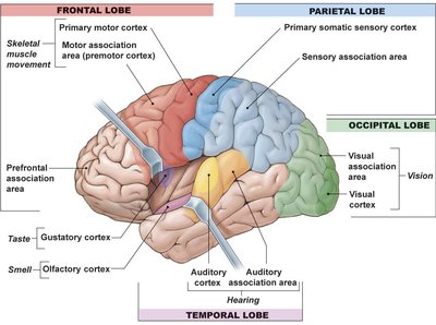
The brain processes sensory information in highly organized regions. Each sensory pathway projects to a specific area of the cerebral cortex, allowing for precise localization and interpretation of stimuli.
The primary somatic sensory cortex receives input from the body’s surface and proprioceptors.
Other sensory modalities (vision, hearing, taste, smell) are processed in distinct cortical regions.
Somatotopic Organization
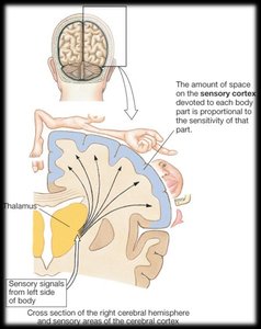
The sensory cortex is organized somatotopically, meaning that different body regions are represented in specific cortical areas. The amount of cortical space devoted to each body part is proportional to its sensory acuity.
Regions with higher sensitivity (e.g., hands, lips) occupy larger cortical areas.
Sensory signals from the left side of the body are processed in the right hemisphere and vice versa.
Somatosensory Receptors
Types and Functions of Skin Receptors
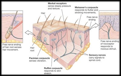
The skin contains various mechanoreceptors, thermoreceptors, and nociceptors, each specialized for detecting different types of stimuli.
Mechanoreceptors: Detect touch, pressure, vibration, and stretch.
Thermoreceptors: Detect changes in temperature.
Nociceptors: Detect potentially damaging stimuli (pain).
Classification of Skin Receptors
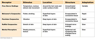
The table below summarizes the main types of skin receptors, their stimuli, locations, structures, and adaptation properties.
Receptor | Stimulus | Location | Structure | Adaptation |
|---|---|---|---|---|
Free Nerve Endings | Temperature, noxious stimuli, hair movement | Around hair roots, under skin surface | Unmyelinated nerve endings | Variable |
Meissner's Corpuscles | Flutter, stroking | Superficial skin layers | Encapsulated in connective tissue | Rapid |
Pacinian Corpuscles | Vibration | Deep skin layers | Encapsulated in connective tissue | Rapid |
Ruffini Corpuscles | Skin stretch | Deep skin layers | Enlarged nerve endings | Slow |
Merkel Receptors | Steady pressure, texture | Epidermal cell synapsing with nerve ending | Enlarged nerve ending | Slow |
Pain Perception and Modulation
Nociception and Pain Types
Pain is the brain’s interpretation of sensory information from nociceptors. It serves as a protective mechanism and is clinically important for diagnosis.
Fast pain: Sharp, well-localized; transmitted by myelinated A𝛿 fibers (~30 m/s).
Slow pain: Dull, diffuse; transmitted by unmyelinated C fibers (~1 m/s).
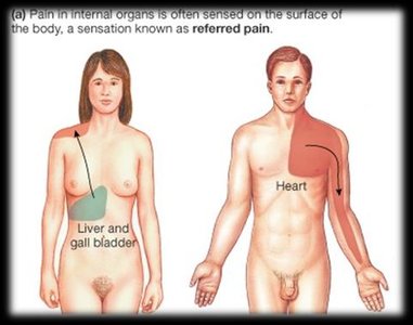
Visceral pain: Originates from internal organs; often poorly localized and may be felt as referred pain.
Referred Pain
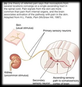
Referred pain occurs when pain from internal organs is perceived as originating from a different, usually superficial, location. This is due to the convergence of visceral and somatic sensory pathways in the spinal cord.
Common example: Cardiac ischemia felt as pain in the left arm or chest.
Pain Modulation: Gate-Control Theory
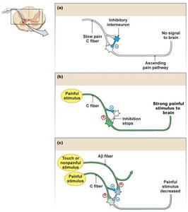
Pain perception can be modulated by other sensory inputs or by descending pathways from the brain. The gate-control theory proposes that non-painful input can inhibit the transmission of pain signals in the spinal cord.
Touch or non-painful stimuli can "close the gate" to painful input, reducing pain perception.
Inhibitory interneurons in the spinal cord play a key role in this process.
Special Senses: Taste and Smell
Gustation (Taste)
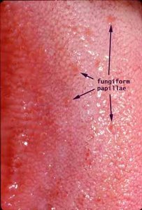
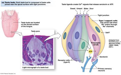
Taste is detected by specialized receptor cells clustered in taste buds, primarily on the tongue but also in other regions of the oral cavity and digestive tract. There are five primary tastes: sweet, sour, salty, bitter, and umami.
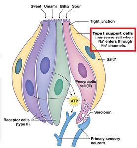
Type I cells: Detect salt via Na+ channels.
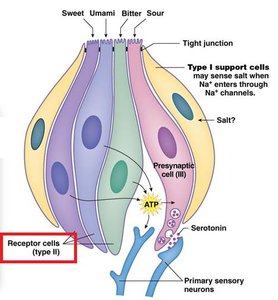
Type II cells: Detect sweet, bitter, and umami via G protein-coupled receptors; release ATP as a signal.
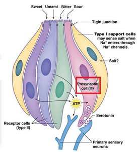
Type III cells: Detect sour; synapse directly with sensory neurons and use serotonin as a neurotransmitter.
Olfaction (Smell)
Olfactory receptor cells are located in the olfactory epithelium of the nasal cavity. Each receptor cell expresses one type of odorant receptor, allowing detection of a wide range of odor molecules. Olfactory neurons are unique in their ability to regenerate throughout life.
Odorant receptors are G protein-coupled receptors (GPCRs).
Olfactory pathways project directly to the olfactory cortex, bypassing the thalamus.
Summary
Sensory physiology encompasses the detection, transmission, and processing of environmental stimuli. Understanding these mechanisms is essential for clinical assessment, diagnosis, and the study of human and animal behavior.
