 Back
BackUnit 3 Study Guide
Study Guide - Smart Notes
 Tailored notes based on your materials, expanded with key definitions, examples, and context.
Sensory Physiology
Overview of Sensory Systems
Sensory physiology explores how the nervous system detects, transmits, and interprets information from the environment. Sensory receptors convert physical or chemical stimuli into electrical signals, which are then processed by the central nervous system to produce perception.
Sensory Receptors: Specialized cells or nerve endings that respond to specific types of stimuli.
Pathways: Sensory information travels from receptors through afferent neurons to the spinal cord and brain.
Perception: The brain interprets sensory signals, allowing us to experience sensations such as touch, pain, temperature, and proprioception.
Types of Sensory Receptors
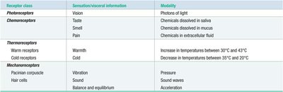
Sensory receptors are classified based on the modality they detect. Each class is specialized for a particular type of stimulus.
Photoreceptors: Detect light for vision.
Chemoreceptors: Respond to chemicals for taste, smell, and pain.
Thermoreceptors: Sense temperature changes.
Mechanoreceptors: Detect mechanical forces such as pressure, vibration, and sound.
General Sensory Pathway
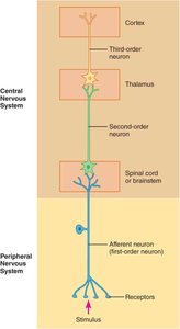
Sensory information is transmitted through a hierarchical pathway involving multiple neurons.
First-order neuron: Afferent neuron that receives input from the receptor.
Second-order neuron: Located in the spinal cord or brainstem; relays information to the thalamus.
Third-order neuron: Projects from the thalamus to the sensory cortex.
Somatosensory System
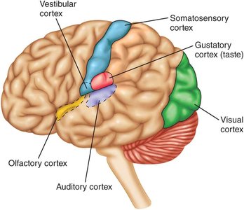
Somatosensory Cortex and Pathways
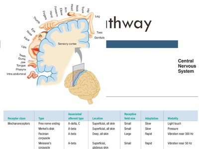
The somatosensory cortex is the primary region for processing touch, proprioception, and pain. Sensory input is mapped onto the cortex, creating a somatotopic representation of the body.
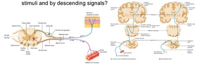
Somatosensory Pathways: Include dorsal column-medial lemniscal and spinothalamic tracts.
Receptive Fields: Each neuron responds to stimuli within a specific area of the skin.
Structure and Function of Sensory Receptors
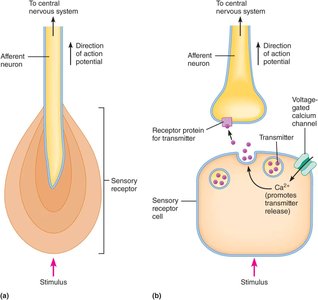
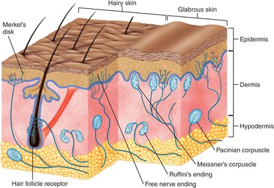
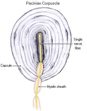
Sensory receptors can be specialized nerve endings or separate cells that synapse onto afferent neurons.
Specialized Endings: Directly generate action potentials in response to stimuli.
Separate Receptor Cells: Release neurotransmitters to activate afferent neurons.
Receptor Potentials and Signal Transmission
Receptor Potentials
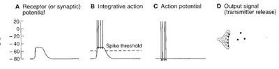
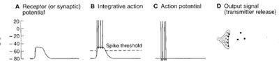
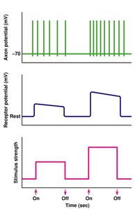
Receptor potentials are graded changes in membrane potential produced by sensory stimuli. If the potential reaches threshold, it triggers action potentials in the afferent neuron.
Graded Response: The amplitude of the receptor potential depends on stimulus strength.
Action Potential Generation: Occurs when the receptor potential exceeds the spike threshold.
Adaptation of Sensory Receptors
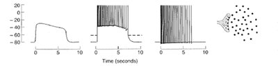
Sensory receptors may adapt to sustained stimuli, altering their response over time.
Slowly Adapting Receptors: Continue to respond throughout the stimulus duration.
Rapidly Adapting Receptors: Respond quickly at stimulus onset and offset.
Stimulus Intensity and Location Coding
Intensity Coding
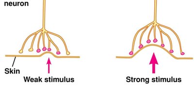
The nervous system codes stimulus intensity by the frequency of action potentials and the number of neurons activated.
Frequency Coding: Stronger stimuli produce higher action potential frequencies.
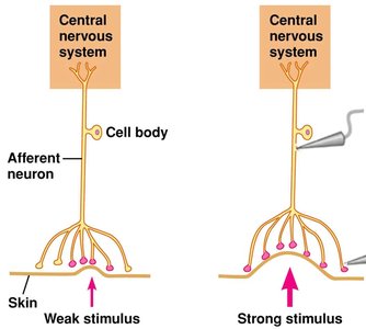
Population Coding: Stronger stimuli recruit more sensory neurons.
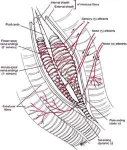
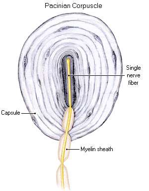
Location Coding
The location of a stimulus is determined by the spatial arrangement of activated neurons and their receptive fields.
Receptive Fields: Each neuron responds to stimuli in a specific area.
Lateral Inhibition: Enhances spatial resolution by inhibiting neighboring neurons, sharpening the perception of stimulus location.
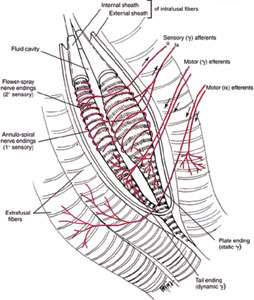

Pain Transmission and Modulation
Nociceptors and Pain Pathways
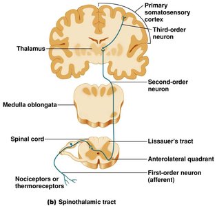
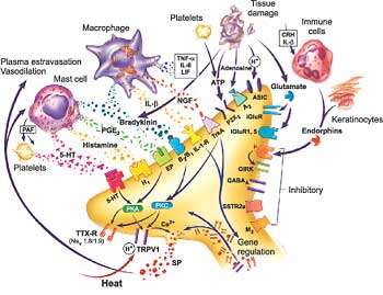
Nociceptors are free nerve endings that detect noxious stimuli, including mechanical, thermal, and chemical signals. Pain signals are transmitted via the spinothalamic pathway to the brain.
Spinothalamic Tract: Carries pain and temperature information from the spinal cord to the thalamus and cortex.
Order of Neurons: First-order (nociceptor), second-order (spinal cord), third-order (thalamus to cortex).
Modulation of Pain
Pain perception can be modified by mechanical stimuli (e.g., touch) and descending signals from the brain.
Gate-Control Theory: Touch input can inhibit pain transmission at the spinal cord level.
Descending Modulation: Brain signals can suppress pain via endogenous analgesia systems.
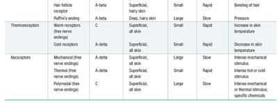
Summary Table: Sensory Receptors in the Skin
Classification and Properties
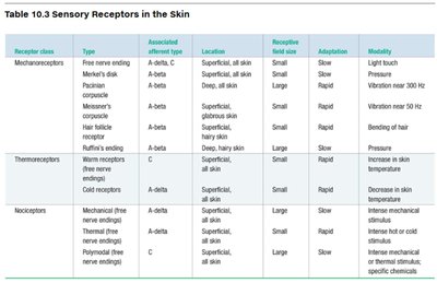
Sensory receptors in the skin are classified by their modality, adaptation, and associated afferent fibers.
Receptor Class | Type | Associated Afferent Type | Location | Receptive Field Size | Adaptation | Modality |
|---|---|---|---|---|---|---|
Mechanoreceptors | Free nerve ending, Merkel's disk, Pacinian corpuscle, Meissner's corpuscle, Hair follicle receptor, Ruffini's ending | A-delta, A-beta | Superficial, all skin; Deep, hairy skin | Small/Large | Rapid/Slow | Light touch, Pressure, Vibration, Bending of hair |
Thermoreceptors | Warm receptors, Cold receptors | A-delta, C | Superficial, all skin | Small | Rapid/Slow | Increase/Decrease in skin temperature |
Nociceptors | Mechanical, Thermal, Polymodal | A-delta, C | Superficial, all skin | Small/Large | Slow | Intense mechanical, thermal, or chemical stimuli |
Key Points and Applications
Sensory coding: Intensity and location are coded by action potential frequency, neuron recruitment, and lateral inhibition.
Pain modulation: Touch and descending signals can alter pain perception.
Clinical relevance: Understanding sensory pathways is essential for diagnosing neurological disorders and managing pain.
Example:
Damage to the dorsal column pathway results in loss of fine touch and proprioception, while pain and temperature sensation remain intact due to the spinothalamic pathway.
Additional info: Academic context was added to clarify receptor types, pathway organization, and coding mechanisms.
