 Back
Sensory Receptors and Spinal Reflexes: Study Guide
Study Guide - Smart Notes
 Tailored notes based on your materials, expanded with key definitions, examples, and context.
Ch. 13b and 15a: Sensory Receptors and Spinal Reflexes

Introduction to Sensation and Perception
Sensation and perception are fundamental processes in the nervous system that allow organisms to detect and interpret changes in their internal and external environments. Sensation refers to the detection of stimuli, while perception is the conscious awareness of these sensations.
General senses: Include detection of temperature, pain, touch, pressure, vibration, and proprioception. These senses use simple receptors distributed throughout the body.
Special senses: Include olfaction, vision, gustation, equilibrium, and hearing. These senses use complex receptors located in specialized sense organs.
Detection of Stimuli
Stimuli are detected by sensory receptors, which convert (transduce) physical or chemical signals into electrical signals (action potentials) that can be interpreted by the nervous system.
Transduction: The process of converting a stimulus into an action potential.
Specificity: Each receptor is sensitive to a particular type of stimulus.
Receptive field: The area monitored by a single receptor; size and distribution vary by region.
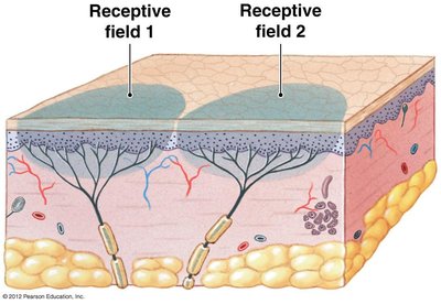
Example: Receptive fields in the skin demonstrate how different areas are monitored by individual sensory neurons, affecting sensitivity and localization of stimuli.
Types of Sensory Receptors
Sensory receptors are classified by the location and type of stimulus they detect.
By location: Exteroceptors (external stimuli), interoceptors (internal stimuli), proprioceptors (body position).
By stimulus type:
Nociceptors: Detect pain from extremes of temperature, mechanical damage, or chemicals.
Thermoreceptors: Detect temperature changes.
Mechanoreceptors: Detect physical distortion (stretch, compression, twisting).
Chemoreceptors: Detect chemical concentrations (e.g., pH, CO2, O2).
Mechanoreceptors and Tactile Receptors
Mechanoreceptors respond to mechanical forces and are subdivided into tactile receptors, baroreceptors, and proprioceptors. Tactile receptors are specialized for touch, pressure, and vibration.
Tactile receptors: Include free nerve endings, root hair plexuses, tactile (Merkel's) discs, tactile (Meissner's) corpuscles, lamellated (Pacinian) corpuscles, and Ruffini corpuscles.
Free nerve endings and tactile discs: Detect touch and pressure in the epidermis; tonic and have small receptive fields.
Root hair plexuses: Detect movement of hair follicles; phasic and adapt quickly.
Tactile corpuscles and lamellated corpuscles: Detect pressure and vibration; complex structures that adapt quickly.
Ruffini corpuscles: Detect deep pressure in the dermis; tonic and intertwined with collagen fibers.
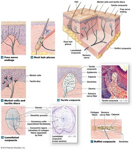
Example: The diagram shows the location and structure of various tactile receptors in the skin, illustrating their roles in detecting different types of mechanical stimuli.
Adaptation of Sensory Receptors
Adaptation is the reduction in sensitivity to a constant stimulus. It can occur at the level of the receptor (peripheral adaptation) or within the central nervous system (central adaptation).
Tonic receptors: Always active; adapt slowly or not at all (e.g., pain receptors).
Phasic receptors: Inactive unless stimulated; adapt quickly (e.g., thermoreceptors).
Neural Circuits and Reflexes
Neuronal pools are groups of CNS interneurons that process sensory information and coordinate responses. Neural circuits describe the flow of information between neurons and pools, including divergence, convergence, serial processing, parallel processing, and reverberation.
Reflexes and Reflex Arcs
Reflexes are rapid, automatic responses to specific stimuli, usually mediated by negative feedback to maintain homeostasis. The reflex arc is the neural circuit connecting a receptor to an effector.
Components of a reflex arc: Sensory neuron, interneuron (optional), motor neuron.
Events in a neural reflex:
Stimulus activates a receptor.
Activation of sensory neuron (graded depolarization causes action potential).
Information processing by interneuron(s).
Activation of motor neuron (action potential travels to periphery).
Activation of peripheral effector.
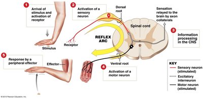
Example: The reflex arc diagram illustrates the pathway from stimulus detection to effector response, highlighting the roles of sensory and motor neurons.
Classification of Reflexes
Reflexes are classified based on development, motor response, circuit complexity, and site of processing.
Development: Innate (genetically programmed) or acquired (learned).
Motor response: Somatic (skeletal muscle) or visceral (autonomic, smooth/cardiac muscle).
Circuit complexity: Monosynaptic (direct synapse) or polysynaptic (interneurons involved).
Routing in spinal cord: Ipsilateral (same side) or contralateral (opposite side).
Processing site: Cranial (brain) or spinal (spinal cord).
Stretch Reflexes
Stretch reflexes automatically regulate skeletal muscle length, such as the patellar tendon (knee-jerk) response. These reflexes help maintain posture and balance.
Muscle spindle: Contains intrafusal (receptor) and extrafusal (effector) muscle fibers.
Reflex arc: Intrafusal fibers → sensory neuron; extrafusal fibers ← motor neuron.
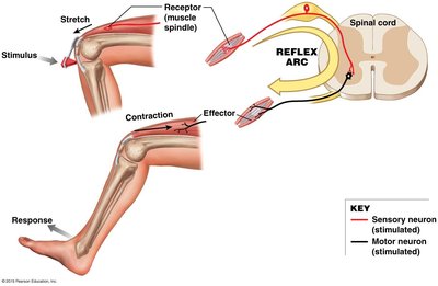
Example: The stretch reflex arc diagram shows the sequence from muscle stretch detection to contraction, maintaining muscle tone and posture.
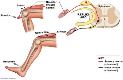
Example: The muscle spindle diagram illustrates the arrangement of intrafusal and extrafusal fibers, and their innervation by sensory and gamma motor neurons.
Withdrawal and Crossed Extensor Reflexes
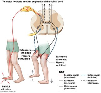
Withdrawal reflexes move body parts away from harmful stimuli, such as pain. The flexor reflex is a common example. The crossed extensor reflex occurs simultaneously, allowing the opposite limb to support body weight.
Flexor reflex: Flexor muscles contract, extensor muscles relax.
Crossed extensor reflex: Extensor muscles stimulated, flexor muscles inhibited on the opposite side.
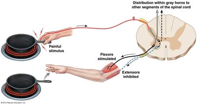
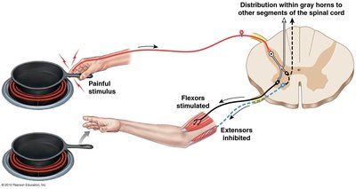
Example: The diagrams show the neural pathways involved in withdrawal and crossed extensor reflexes, demonstrating coordinated responses to painful stimuli.
IV. Integration and Control of Spinal Reflexes
Effects of Higher CNS Centers
Higher centers in the brain can modulate spinal reflexes through facilitation (enhancement) or inhibition (suppression). This allows for conscious control and fine-tuning of reflex responses.
Facilitation: Enhances reflexes (e.g., reinforcement during heightened alertness).
Inhibition: Suppresses reflexes, either chronically (e.g., negative Babinski reflex) or temporarily by conscious effort.

Example: The plantar reflex (negative Babinski) is normal in adults, while the Babinski sign (positive Babinski) is normal in infants but pathological in adults, indicating loss of descending inhibition.
Descending Pathways and Reflex Modulation
Descending pathways from the brain can activate neuronal pools responsible for reflex motor patterns, allowing for facilitation and inhibition of ongoing motions.
Fine-tuning: The brain can adjust reflex responses to suit complex motor tasks.
Summary Table: Types of Sensory Receptors
Receptor Type | Stimulus Detected | Location | Adaptation |
|---|---|---|---|
Nociceptors | Pain (temperature, mechanical, chemical) | Skin, joints, periosteum, blood vessels | Tonic; do not adapt |
Thermoreceptors | Temperature | Dermis, muscles, liver, hypothalamus | Phasic; adapt quickly |
Mechanoreceptors | Physical distortion | Skin, muscles, organs | Varies |
Chemoreceptors | Chemical concentration | Blood, CSF | Adapt |
Summary Table: Types of Reflexes
Classification | Type | Description |
|---|---|---|
Development | Innate | Genetically programmed |
Development | Acquired | Learned |
Motor Response | Somatic | Skeletal muscle |
Motor Response | Visceral | Autonomic (smooth/cardiac muscle) |
Circuit Complexity | Monosynaptic | Direct synapse |
Circuit Complexity | Polysynaptic | Interneurons involved |
Routing | Ipsilateral | Same side |
Routing | Contralateral | Opposite side |
Processing Site | Cranial | Brain |
Processing Site | Spinal | Spinal cord |
Additional info: Academic context was added to clarify definitions, examples, and the functional significance of sensory receptors and reflexes. Tables were inferred for summary and classification purposes.
