 Back
BackSkeletal and Smooth Muscle: Structure, Function, and Contraction Mechanisms
Study Guide - Smart Notes
 Tailored notes based on your materials, expanded with key definitions, examples, and context.
Overview of Muscle Types
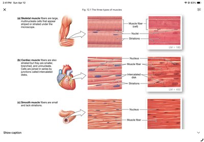
Three Types of Muscle
Muscle tissue in the human body is classified into three main types, each with distinct structure and function:
Skeletal Muscle: Striated muscle attached to bones, responsible for voluntary body movement. Controlled by somatic motor neurons.
Cardiac Muscle: Striated muscle found only in the heart, responsible for pumping blood. Involuntary control via autonomic innervation and the endocrine system.
Smooth Muscle: Non-striated muscle found in walls of internal organs and tubes, controlling movement of materials within the body. Involuntary control via autonomic innervation and the endocrine system.
Skeletal Muscle Structure and Organization
Attachment and Function
Skeletal muscles are typically attached to bones by tendons and are responsible for moving the skeleton:
Origin: The attachment site closest to the trunk or more stationary bone.
Insertion: The more distal or more mobile attachment.
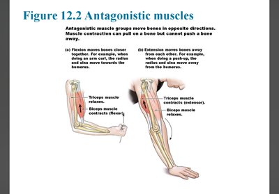
Flexor: Muscle that brings bones together, decreasing joint angle.
Extensor: Muscle that moves bones away, increasing joint angle.
Flexor-extensor pairs form antagonistic muscle groups.
Muscle Fiber Structure
Skeletal muscle fibers are long, cylindrical cells with multiple nuclei. Key structural features include:
Muscle fibers: The actual muscle cells, bundled into fascicles.
Satellite cells: Stem cells involved in muscle regeneration and repair.
Connective tissue sheaths: Surround individual fibers and the entire muscle, forming tendons.
Muscle Fiber Anatomy
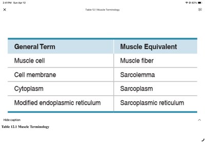
Muscle fibers have specialized terminology for their structures:
General Term | Muscle Equivalent |
|---|---|
Muscle cell | Muscle fiber |
Cell membrane | Sarcolemma |
Cytoplasm | Sarcoplasm |
Modified endoplasmic reticulum | Sarcoplasmic reticulum |
Myofibrils: Contractile structures within muscle fibers.
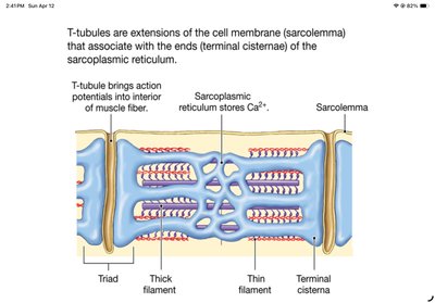
Transverse tubules (T-tubules): Extensions of the sarcolemma that allow action potentials to penetrate the fiber's interior.
Sarcoplasmic reticulum: Stores and releases Ca2+ for contraction.
Glycogen granules and mitochondria provide energy for contraction.
Myofibril Structure and Sarcomere Organization
Contractile and Accessory Proteins
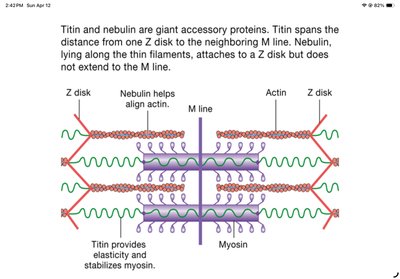
Thin filament: Composed of actin (contractile protein), tropomyosin and troponin (regulatory proteins), and titin and nebulin (accessory proteins).
Thick filament: Composed of myosin (contractile protein) with ATPase activity, forming crossbridges with actin.
Sarcomere Structure
The sarcomere is the functional contractile unit of the myofibril:
Z disks: Define the boundaries of a sarcomere; thin filaments attach here.
I band: Contains only thin filaments.
A band: Contains regions of overlapping thick and thin filaments.
H zone: Central region with only thick filaments.
M line: Proteins to which thick filaments attach.
Accessory Proteins
Titin: Elastic protein that stabilizes thick filaments and returns stretched muscles to resting length.
Nebulin: Aligns and stabilizes thin filaments.
Muscle Contraction Mechanisms
Muscle Tension and Contraction
Muscle tension: Force created by muscle contraction.
Load: Force opposing contraction.
Contraction: Creation of tension in muscle.
Relaxation: Release of tension.
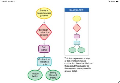
Major steps in skeletal muscle contraction:
Events at the neuromuscular junction (NMJ)
Excitation-contraction (E-C) coupling
Contraction-relaxation cycle
Events at the Neuromuscular Junction and E-C Coupling
Acetylcholine (ACh) is released from the somatic motor neuron.
ACh binds to receptors on the sarcolemma, causing Na+ influx and K+ efflux, leading to depolarization (end-plate potential) and muscle action potential.
The action potential triggers Ca2+ release from the sarcoplasmic reticulum via DHP and RyR receptors.
Calcium binds to troponin, initiating contraction.
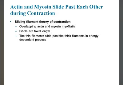
Sliding Filament Theory of Contraction
Muscle contraction occurs as thin filaments slide past thick filaments, shortening the sarcomere without changing the length of the filaments themselves. This process is energy-dependent and underlies all muscle contraction.
Calcium Signals and Contraction
Troponin: Binds Ca2+ and controls the position of tropomyosin.
Tropomyosin: Blocks myosin-binding sites on actin at rest.
When Ca2+ binds troponin, tropomyosin moves, exposing binding sites for myosin on actin, allowing contraction to proceed.
Myosin-Actin Crossbridge Cycle
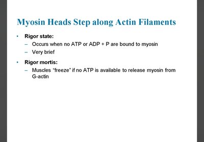
Rigor state: Myosin tightly bound to actin after a power stroke.
ATP binds myosin, causing detachment from actin.
ATP hydrolysis "cocks" the myosin head.
Myosin binds new actin site, releases Pi, and performs the power stroke.
ADP is released, and the cycle repeats as long as ATP and Ca2+ are available.
Rigor mortis occurs when ATP is depleted, causing muscles to remain contracted.
Muscle Metabolism and Fiber Types
ATP Supply for Contraction
Phosphocreatine: Provides a rapid, short-term source of ATP via creatine kinase.
Anaerobic glycolysis: Quick ATP production without oxygen, but yields less energy and produces lactate.
Aerobic respiration: Slower, oxygen-dependent, but yields much more ATP.
Classification of Skeletal Muscle Fibers
Slow-twitch fibers (Type I): Rely on oxidative phosphorylation, have high myoglobin, mitochondria, and capillaries; fatigue-resistant.
Fast-twitch fibers (Type IIB/IIX): Rely on glycolysis, fatigue quickly, generate rapid, powerful contractions.
Fast-oxidative-glycolytic fibers (Type IIA): Use both oxidative and glycolytic metabolism; intermediate properties.
Muscle Contraction Regulation
All-or-None Principle and Graded Contractions
A single muscle fiber contracts fully if threshold is reached (all-or-none law).
Whole muscle force is graded by recruiting more fibers and varying stimulation frequency.
Resting Fiber Length and Tension
Optimal sarcomere length produces maximal tension; too short or too long reduces force.
Summation: Increased contraction force when stimuli arrive before full relaxation.
Tetanus: Maximal, sustained contraction with little or no relaxation.
Motor unit: One motor neuron and all the muscle fibers it innervates; recruitment increases contraction force.
