 Back
Skeletal Muscle Movements, Roles, and Anatomy: ANP College Study Guide
Study Guide - Smart Notes
 Tailored notes based on your materials, expanded with key definitions, examples, and context.
Muscle Movements, Roles, and Names
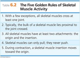
The Five Golden Rules of Skeletal Muscle Activity
Understanding skeletal muscle activity is fundamental to anatomy and physiology. The following rules summarize the basic principles governing muscle function:
Rule 1: With few exceptions, all skeletal muscles cross at least one joint.
Rule 2: The bulk of a skeletal muscle usually lies proximal to the joint it crosses.
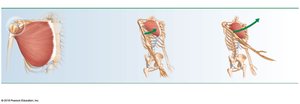
Rule 3: All skeletal muscles have at least two attachments: the origin (fixed) and the insertion (movable).
Rule 4: Skeletal muscles can only pull; they never push.
Rule 5: During contraction, the muscle insertion moves toward the origin.
Types of Body Movements
Muscle Attachments: Origin and Insertion
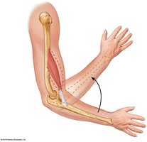
Muscles are attached to bones at two points: the origin (immovable or less movable) and the insertion (movable). When a muscle contracts, the insertion moves toward the origin, producing movement at the joint.
Flexion, Extension, and Hyperextension
These are fundamental movements at joints:
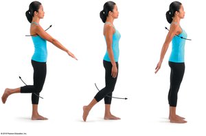
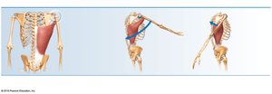
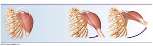
Flexion: Decreases the angle between two bones, bringing them closer together. Common in hinge joints (e.g., knee, elbow) and ball-and-socket joints (e.g., hip).
Extension: Increases the angle between two bones, moving them farther apart. Opposite of flexion.
Hyperextension: Extension beyond the normal range (180º).
Rotation
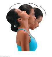
Rotation is the movement of a bone around its longitudinal axis, common in ball-and-socket joints. Example: shaking your head "no" involves rotating the atlas around the dens of the axis.
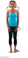
Abduction, Adduction, and Circumduction
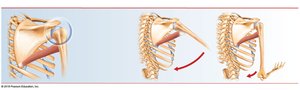
Abduction: Movement of a limb away from the midline.
Adduction: Movement of a limb toward the midline.
Circumduction: Combination of flexion, extension, abduction, and adduction; distal end moves in a circle while proximal end remains stationary.
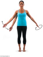
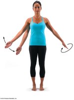
Special Movements
Dorsiflexion: Lifting the foot so the superior surface approaches the shin.
Plantar flexion: Pointing the toes away from the head.
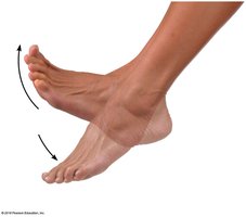
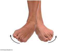
Inversion: Turning the sole of the foot medially.
Eversion: Turning the sole of the foot laterally.
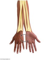
Supination: Forearm rotates laterally so palm faces anteriorly; radius and ulna are parallel.
Pronation: Forearm rotates medially so palm faces posteriorly; radius and ulna cross.
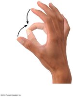
Opposition: Moving the thumb to touch the tips of other fingers on the same hand.
Interactions of Skeletal Muscles in the Body
Muscle Action and Coordination
Skeletal muscles work in groups to produce coordinated movements. Muscles can only pull as they contract, not push. Groups producing opposite actions are located on opposite sides of a joint.
Muscle Roles
Prime mover: Muscle with major responsibility for a movement.
Antagonist: Muscle that opposes or reverses a prime mover.
Synergist: Muscle that aids a prime mover or reduces undesirable movements.
Fixator: Specialized synergists that stabilize the origin of a prime mover.
Muscle Action Relative to Joint Position
Anterior side: Produces flexion (except knee and ankle).
Posterior side: Produces extension (except knee and ankle).
Lateral side: Produces abduction.
Medial side: Produces adduction.
Naming Skeletal Muscles
Criteria for Muscle Naming
Muscles are named based on several criteria:
Direction of fibers: e.g., rectus (straight)
Relative size: e.g., maximus (largest)
Location: e.g., temporalis (temporal bone)
Number of origins: e.g., triceps (three heads)
Origin and insertion: e.g., sternocleidomastoid (originates on sternum and clavicle, inserts on mastoid process)
Shape: e.g., deltoid (triangular)
Action: e.g., flexor and extensor (flexes or extends a bone)
Fascicle Arrangement and Muscle Structure
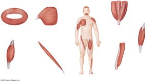
The arrangement of muscle fascicles affects muscle shape and function. Types include circular, convergent, parallel, fusiform, unipennate, bipennate, and multipennate.
Superficial Muscles of the Body
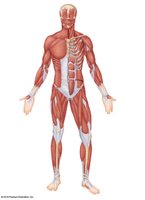
Superficial Anterior Muscles
Major muscles of the head, neck, trunk, arm, and leg are classified by their origin, insertion, and primary action.
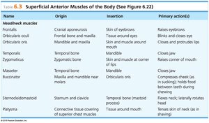
Name | Origin | Insertion | Primary action(s) |
|---|---|---|---|
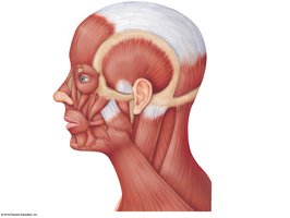
Frontalis | Cranial aponeurosis | Skin of eyebrows | Raises eyebrows |
Orbicularis oculi | Frontal bone and maxilla | Tissue around eyes | Blinks and closes eyes |
Orbicularis oris | Mandible and maxilla | Skin and muscle around mouth | Closes and protrudes lips |
Temporalis | Temporal bone | Mandible | Closes jaw |
Zygomaticus | Zygomatic bone | Skin and muscle at corner of mouth | Raises corner of mouth |
Masseter | Temporal bone | Mandible | Closes jaw |
Buccinator | Maxilla and mandible near molars | Orbicularis oris | Compresses cheek (as in sucking); holds food between teeth during chewing |
Sternocleidomastoid | Sternum and clavicle | Temporal bone (mastoid process) | Flexes neck; laterally rotates head |
Platysma | Connective tissue covering of superior chest muscles | Tissue around mouth | Pulls corners of mouth inferiorly |
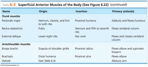
Name | Origin | Insertion | Primary action(s) |
|---|---|---|---|
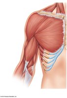
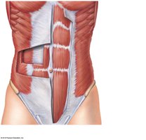
Pectoralis major | Sternum, clavicle, and first to sixth ribs | Proximal humerus | Adducts and flexes humerus |
Rectus abdominis | Pubis | Sternum and fifth to seventh ribs | Flexes vertebral column |
External oblique | Lower eight ribs | Iliac crest | Flexes and rotates vertebral column |
Biceps brachii | Scapula of shoulder girdle | Proximal radius | Flexes elbow and supinates forearm |
Brachialis | Distal humerus | Proximal ulna | Flexes elbow |
Deltoid | See Table 6.4 | Abducts arm |
Name | Origin | Insertion | Primary action(s) |
|---|---|---|---|
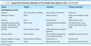
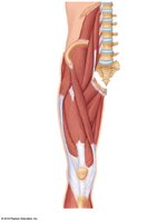
Iliopsoas | Ilium and lumbar vertebrae | Femur (lesser trochanter) | Flexes hip |
Adductor muscles | Pelvis | Proximal femur | Adduct and medially rotate thigh |
Sartorius | Ilium | Proximal tibia | Flexes thigh on hip |
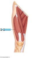
Quadriceps group | Vasti: femur; rectus femoris: pelvis | Tibial tuberosity via patellar ligament | All extend knee; rectus femoris also flexes hip on thigh |
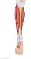
Tibialis anterior | Proximal tibia | First cuneiform (tarsal) and first metatarsal of foot | Dorsiflexes and inverts foot |
Extensor digitorum longus | Proximal tibia and fibula | Distal toes 2–5 | Extends toes |
Fibularis muscles | Fibula | Metatarsals of foot | Plantar flex and evert foot |
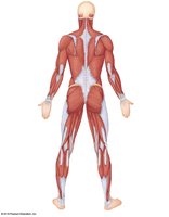
Superficial Posterior Muscles
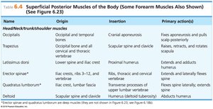
Name | Origin | Insertion | Primary action(s) |
|---|---|---|---|
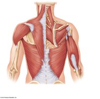
Occipitalis | Occipital and temporal bones | Cranial aponeurosis | Pulls scalp posteriorly |
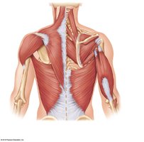
Trapezius | Occipital bone and all cervical and thoracic vertebrae | Scapular spine and clavicle | Raises, retracts, and rotates scapula |
Latissimus dorsi | Lower spine and iliac crest | Proximal humerus | Extends and adducts humerus |
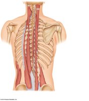
Erector spinae | Iliac crests, ribs 3–12, and vertebrae | Ribs, thoracic and cervical vertebrae | Extends and laterally flexes spine |
Quadratus lumborum | Iliac crest, lumbar fascia | Transverse processes of lumbar vertebrae | Flexes spine laterally |
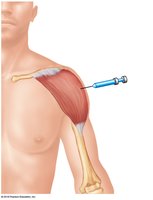
Deltoid | Scapular spine and clavicle | Humerus (deltoid tuberosity) | Abducts humerus |
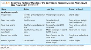
Name | Origin | Insertion | Primary action(s) |
|---|---|---|---|
Triceps brachii | Shoulder girdle and proximal humerus | Olecranon process of ulna | Extends elbow |
Flexor carpi radialis | Distal humerus | Second and third metacarpals | Flexes wrist and abducts hand |
Flexor carpi ulnaris | Distal humerus and posterior ulna | Carpals of wrist and fifth metacarpal | Flexes wrist and adducts hand |
Extensor carpi radialis | Humerus | Second and third metacarpals | Extends wrist and abducts hand |
Extensor digitorum | Distal humerus | Base of second and third phalanges of fingers 2–5 | Extends fingers |
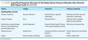
Name | Origin | Insertion | Primary action(s) |
|---|---|---|---|
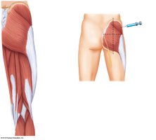
Gluteus maximus | Sacrum and ilium | Proximal femur (gluteal tuberosity) | Extends hip (when forceful extension is required) |
Gluteus medius | Ilium | Proximal femur | Abducts thigh; steadies pelvis during walking |
Hamstring group (biceps femoris, semimembranosus, semitendinosus) | Ischial tuberosity | Proximal tibia (head of fibula in case of biceps femoris) | Flexes knee and extends hip |
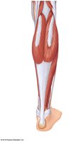
Gastrocnemius | Distal femur | Calcaneus (heel via calcaneal tendon) | Plantar flexes foot and flexes knee |
Soleus | Proximal tibia and fibula | Calcaneus | Plantar flexes foot |
Developmental Aspects of the Muscular System
Muscle Control and Nervous System Maturation
Increasing muscular control reflects the maturation of the nervous system. Muscle control is achieved in a superior/inferior and proximal/distal direction.
Exercise and Muscle Health
Regular exercise is essential for muscle health.
Without exercise, muscles atrophy (decrease in size).
With vigorous exercise, muscles hypertrophy (increase in size).
Aging and Muscle Mass
As we age, muscle mass decreases and muscles become more sinewy.
Exercise helps retain muscle mass and strength.
Example: Elderly individuals who exercise regularly maintain greater muscle strength and mass compared to those who are sedentary.
