 Back
BackSkeletal Muscle Physiology: Structure, Function, and Excitation-Contraction Coupling
Study Guide - Smart Notes
 Tailored notes based on your materials, expanded with key definitions, examples, and context.
Skeletal Muscle Structure and Function
Overview of Skeletal Muscle Anatomy
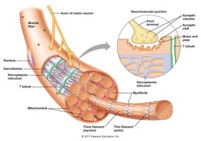
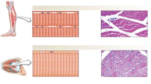
Skeletal muscle is composed of long, multinucleated fibers that are responsible for voluntary movements. Each muscle fiber contains myofibrils, which are made up of repeating units called sarcomeres—the fundamental contractile units of muscle.
Muscle Fiber: A single muscle cell, formed by the fusion of multiple myocytes during development.
Myofibrils: Cylindrical structures within muscle fibers, composed of sarcomeres in series.
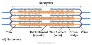
Sarcomere: The basic contractile unit, defined by the region between two Z lines. Contains thick (myosin) and thin (actin) filaments.
Striations: The banding pattern seen in skeletal muscle, resulting from the ordered arrangement of sarcomeres.
Muscle Contraction and Movement
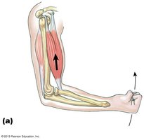
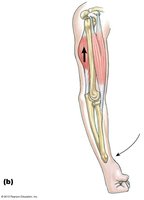
Muscle contraction results from the sliding of thick and thin filaments within the sarcomere, leading to shortening of the muscle and movement of bones at joints.
Flexion and Extension: Muscles work in antagonistic pairs to produce movement. For example, contraction of the biceps brachii flexes the forearm, while contraction of the triceps brachii extends it.
Neuromuscular Junction and Excitation
Structure and Function of the Neuromuscular Junction (NMJ)
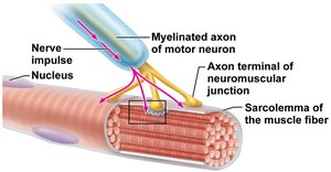
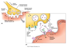
The neuromuscular junction is the synapse between a motor neuron and a skeletal muscle fiber. It is the site where nerve impulses are transmitted to muscle fibers to initiate contraction.
Motor Neuron: Sends action potentials to the muscle fiber.
Axon Terminal: Releases the neurotransmitter acetylcholine (ACh) into the synaptic cleft.
Sarcolemma: The plasma membrane of the muscle fiber, which contains receptors for ACh.
Mechanism of Synaptic Transmission
When an action potential reaches the axon terminal, ACh is released and binds to nicotinic acetylcholine receptors (nAChRs) on the muscle fiber, causing depolarization and initiation of a muscle action potential.
ACh Release: Triggered by Ca2+ influx into the axon terminal.
nAChR: A ligand-gated ion channel that allows Na+ influx, depolarizing the muscle membrane.
AChE (Acetylcholinesterase): Enzyme that rapidly degrades ACh, terminating the signal.
Excitation-Contraction Coupling
From Action Potential to Muscle Contraction
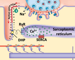
Excitation-contraction (EC) coupling is the process by which an action potential in the muscle fiber leads to contraction. This involves the release of Ca2+ from the sarcoplasmic reticulum (SR) and its interaction with the contractile machinery.
T-tubules: Invaginations of the sarcolemma that transmit the action potential deep into the muscle fiber.
DHP Receptor: Voltage-sensitive L-type Ca2+ channel in the T-tubule membrane.
RyR (Ryanodine Receptor): Ca2+ release channel in the SR membrane, physically coupled to the DHP receptor.
SERCA Pump: Actively transports Ca2+ back into the SR, terminating contraction.
Sarcomere Structure and the Sliding Filament Model
Organization of Thick and Thin Filaments
The sarcomere contains overlapping thick (myosin) and thin (actin) filaments. The arrangement of these filaments is responsible for muscle contraction through the sliding filament mechanism.
Thick Filaments: Composed of myosin, which has ATPase activity and forms cross-bridges with actin.
Thin Filaments: Composed of actin, tropomyosin, and troponin.
Titin: A large protein that maintains the alignment of thick and thin filaments and provides elasticity.

Sliding Filament Theory
During contraction, myosin heads bind to actin and pull the thin filaments toward the center of the sarcomere, shortening the sarcomere without changing the length of the filaments themselves.
Cross-Bridge Cycle: The process by which myosin heads attach to, pivot, and detach from actin, powered by ATP hydrolysis.
Role of Ca2+: Ca2+ binds to troponin, causing tropomyosin to move and expose myosin-binding sites on actin.

Muscle Fiber Types and Energy Metabolism
Types of Skeletal Muscle Fibers
Skeletal muscles contain different fiber types, each adapted for specific functions based on their metabolic and contractile properties.
Property | Slow Oxidative (Type I) | Fast Oxidative (Type IIa) | Fast Glycolytic (Type IIx) |
|---|---|---|---|
Oxidative Capacity | High | High | Low |
Glycolytic Capacity | Low | Intermediate | High |
Speed of Contraction | Low | Intermediate | High |
Myosin ATPase Activity | Low | High | High |
Fatigue Resistance | High | Intermediate | Low |
Fiber Diameter | Small | Intermediate | Large |
Force-Generating Capacity | Small | Intermediate | Large |
ATP Regeneration in Muscle
Muscle contraction requires a continuous supply of ATP, which is regenerated by several mechanisms:
Creatine Phosphate: Provides a rapid but short-lived source of ATP.
Anaerobic Glycolysis: Produces ATP quickly but less efficiently, resulting in lactic acid formation.
Aerobic Respiration: Generates ATP more slowly but can sustain contraction for longer periods.
Motor Units and Control of Muscle Force
Motor Unit Structure and Recruitment
A motor unit consists of a single motor neuron and all the muscle fibers it innervates. The number and size of motor units determine the precision and strength of muscle contraction.
Small Motor Units: Allow fine control (e.g., in the fingers).
Large Motor Units: Generate more force but less precise control (e.g., in the quadriceps).
Size Principle: Smaller motor units are recruited before larger ones as force requirements increase.
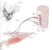
Neural Control of Muscle Contraction
The central nervous system regulates muscle force by varying the number of active motor units and the frequency of action potentials.
Summation: Increased frequency of action potentials leads to greater force production.
Reflex Arcs: Simple neural circuits that mediate automatic responses to stimuli.
Key Equations and Concepts
Ohm’s Law (for neurons):
Ion Current:
Creatine Kinase Reaction:
Summary
Skeletal muscle contraction is a highly regulated process involving the integration of neural signals, excitation-contraction coupling, and the coordinated action of contractile proteins within the sarcomere. The diversity of muscle fiber types and the organization of motor units allow for a wide range of movements and force generation, essential for daily activities and complex motor tasks.
