 Back
BackSkeletal Muscle Structure and Neuromuscular Junction: Study Notes
Study Guide - Smart Notes
 Tailored notes based on your materials, expanded with key definitions, examples, and context.
Muscle Tissue
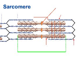
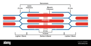
Structure of the Sarcomere
The sarcomere is the fundamental contractile unit of striated muscle fibers. It is defined as the segment between two Z lines and contains organized arrays of actin (thin) and myosin (thick) filaments, which interact to produce muscle contraction.
Z Line: Defines the boundary of each sarcomere and anchors the thin filaments.
Actin Filaments (Thin Filaments): Extend from the Z line toward the center of the sarcomere.
Myosin Filaments (Thick Filaments): Located in the center of the sarcomere, overlapping with actin filaments.
A Band: The region containing the entire length of the thick filaments; appears dark under the microscope.
I Band: The region containing only thin filaments; appears lighter.
H Zone: The central region of the A band where only thick filaments are present (no overlap with thin filaments).
M Line: The center of the sarcomere, where thick filaments are linked by accessory proteins.
Actin and Myosin Filaments
Actin and myosin are the primary proteins responsible for muscle contraction. Their interaction is regulated by accessory proteins and the presence of calcium ions.
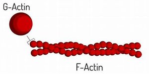
G-Actin: Globular actin monomers that polymerize to form F-actin.
F-Actin: Filamentous actin, a double helical polymer of G-actin subunits.
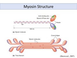
Myosin: Thick filament protein with a tail and two heads, each containing an actin-binding site and ATPase activity.
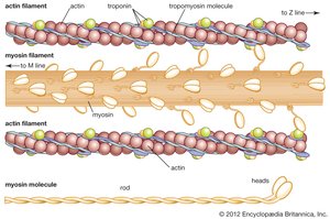
Regulatory Proteins: Troponin and Tropomyosin
Muscle contraction is regulated by the proteins troponin and tropomyosin, which control the exposure of myosin-binding sites on actin filaments.
Tropomyosin: A long, fibrous protein that covers myosin-binding sites on actin in resting muscle.
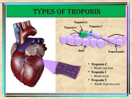
Troponin: A complex of three subunits:
Troponin C (TnC): Binds calcium ions.
Troponin I (TnI): Inhibits actin-myosin interaction.
Troponin T (TnT): Binds to tropomyosin, anchoring the troponin complex.
Role of Calcium in Muscle Contraction
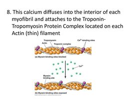
Calcium ions play a critical role in initiating muscle contraction by binding to troponin C, causing a conformational change that moves tropomyosin away from the myosin-binding sites on actin.
When calcium binds to troponin C, the inhibition by troponin I is relieved, and myosin heads can bind to actin, initiating contraction.
Removal of calcium returns the muscle to a relaxed state.
Length-Tension Relationship
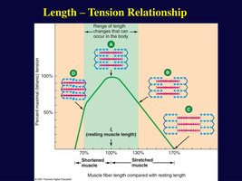
The length-tension relationship describes how the force a muscle fiber can generate depends on its length at the time of stimulation. Maximum tension is produced when there is optimal overlap between actin and myosin filaments.
Too much or too little overlap results in decreased force production.
This relationship is important for understanding muscle function and efficiency.
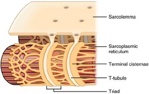
Sarcoplasmic Reticulum and T-Tubules
The sarcoplasmic reticulum (SR) is a specialized endoplasmic reticulum in muscle cells that stores calcium ions. T-tubules are invaginations of the sarcolemma that help transmit action potentials deep into the muscle fiber, ensuring coordinated contraction.
Terminal cisternae: Enlarged areas of the SR adjacent to T-tubules; together they form a triad.
Triad: Structure formed by a T-tubule and two terminal cisternae, critical for excitation-contraction coupling.
Neuromuscular Junction and Muscle Activation
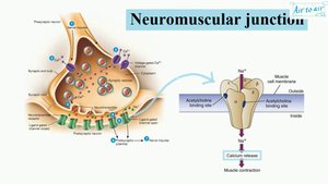
Neuromuscular Junction (NMJ)
The neuromuscular junction is the synapse between a motor neuron and a skeletal muscle fiber. It is the site where nerve impulses are transmitted to initiate muscle contraction.
Motor neuron axon terminals release the neurotransmitter acetylcholine (ACh) into the synaptic cleft.
ACh binds to nicotinic acetylcholine receptors on the muscle cell membrane, leading to depolarization and initiation of an action potential in the muscle fiber.
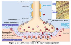
Nicotinic Acetylcholine Receptor (nAChR)
The nicotinic acetylcholine receptor is a pentameric ligand-gated ion channel composed of five subunits (α, β, γ, δ, and ε in adults). It mediates the effects of acetylcholine at the NMJ.
Binding of ACh opens the channel, allowing Na+ influx and K+ efflux, resulting in depolarization of the muscle membrane.
This depolarization triggers an action potential that spreads along the sarcolemma and into the T-tubules.
Excitation-Contraction Coupling
Excitation-contraction coupling refers to the sequence of events by which an action potential in the muscle fiber leads to contraction.
Action potential travels along the sarcolemma and down T-tubules.
This triggers the release of Ca2+ from the sarcoplasmic reticulum.
Ca2+ binds to troponin, initiating contraction as described above.
Summary Table: Key Proteins and Structures in Muscle Contraction
Component | Function |
|---|---|
Actin (Thin Filament) | Provides binding sites for myosin; forms the backbone of the thin filament |
Myosin (Thick Filament) | Motor protein that binds to actin and hydrolyzes ATP for contraction |
Tropomyosin | Covers myosin-binding sites on actin in resting muscle |
Troponin | Regulates position of tropomyosin; binds Ca2+ |
Sarcoplasmic Reticulum | Stores and releases Ca2+ for contraction |
T-tubule | Conducts action potentials into muscle fiber |
nAChR | Ligand-gated ion channel for ACh at NMJ |
Additional info:
The notes above integrate foundational concepts from muscle tissue structure, the molecular basis of contraction, and the physiology of the neuromuscular junction, all of which are core topics in college-level anatomy and physiology courses.
