 Back
Skeletal Muscle System Physiology: Structure, Function, and Mechanisms
Study Guide - Smart Notes
 Tailored notes based on your materials, expanded with key definitions, examples, and context.
Skeletal Muscle System Physiology
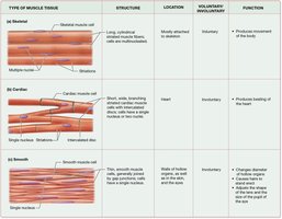
Types of Muscle Tissue
Muscle tissue is classified into three main types, each with distinct structural and functional characteristics. Understanding these differences is essential for grasping their roles in the human body.
Skeletal Muscle: Long, cylindrical, multinucleated cells; striated; voluntary control; attached to bones; responsible for movement.
Cardiac Muscle: Short, branched cells; single nucleus; striated; involuntary control; found in the heart; responsible for pumping blood.
Smooth Muscle: Spindle-shaped cells; single nucleus; non-striated; involuntary control; found in walls of hollow organs; responsible for regulating internal movements.
Type | Structure | Location | Voluntary/Involuntary | Function |
|---|---|---|---|---|
Skeletal | Long, cylindrical, striated, multinucleated | Attached to bones | Voluntary | Movement of skeleton |
Cardiac | Short, branched, striated, single nucleus | Heart | Involuntary | Pumping blood |
Smooth | Spindle-shaped, non-striated, single nucleus | Walls of hollow organs | Involuntary | Regulates internal movements |
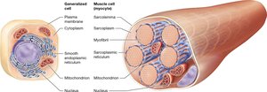
Basic Anatomical Organization of Skeletal Muscle
Skeletal muscle is organized hierarchically from the whole muscle down to the molecular level. This structure is essential for its function in force generation and movement.
Whole muscle: Organ composed of many bundles of muscle fibers.
Muscle fiber: Single, long, multinucleated muscle cell.
Myofibril: Cylindrical contractile structures within each muscle fiber.
Sarcomere: Repeating functional unit of a myofibril; contains actin and myosin, responsible for contraction.
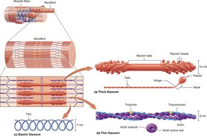
Structure and Function of Muscle Proteins
Muscle contraction relies on the interaction of contractile and regulatory proteins within the sarcomere.
Contractile Proteins:
Actin: Thin filament; provides binding sites for myosin during contraction.
Myosin: Thick filament; motor protein with heads that bind actin and generate force.
Regulatory Proteins:
Troponin: Binds calcium and moves tropomyosin.
Tropomyosin: Blocks myosin-binding sites on actin at rest.
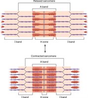
Thin and Thick Filaments in a Sarcomere
The sarcomere is the fundamental contractile unit of muscle, where thin (actin) and thick (myosin) filaments interact. Muscle contraction occurs as myosin pulls actin toward the center, shortening the sarcomere.
Filament overlap: Changes during contraction, increasing force generation.
Sarcomere shortening: Filaments slide past each other; their length does not change.
Neuromuscular Junction and Excitation-Contraction Coupling
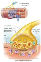
The neuromuscular junction (NMJ) is the site where a motor neuron communicates with a muscle fiber, initiating contraction. Excitation-contraction coupling links the electrical signal to mechanical contraction.
Motor neuron: Releases acetylcholine (ACh) to stimulate the muscle.
Motor end plate: Specialized region of the muscle fiber membrane.
Receptor type: Ligand-gated ACh receptors.
Effector cell: Skeletal muscle fiber.
Roles of Muscle Fiber Structures
Several specialized structures within the muscle fiber facilitate contraction.
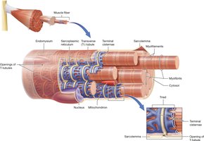
Sarcolemma: Muscle fiber cell membrane; conducts action potentials.
T-tubules: Extensions of the sarcolemma; carry action potential deep into the cell.
Sarcoplasmic reticulum (SR): Specialized smooth ER; stores and releases calcium.
Generation of Resting Membrane Potential in Skeletal Muscle Fiber
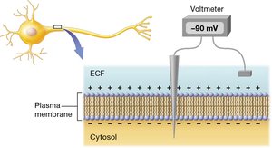
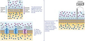
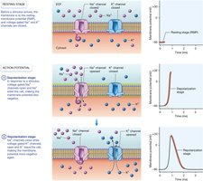
The resting membrane potential is a voltage difference across the sarcolemma, essential for muscle excitability.
Typical value: Approximately -90 mV.
Maintained by: Ion gradients (Na+, K+) and selective permeability.
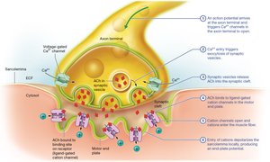
Motor End Plate Excitation and Muscle Action Potential
Excitation at the motor end plate leads to the generation of a muscle action potential, which triggers contraction.
Action potential arrives at motor neuron axon terminal.
Voltage-gated Ca2+ channels open; Ca2+ enters neuron.
ACh released into synaptic cleft.
ACh binds ligand-gated receptors on motor end plate.
Na+ enters muscle fiber, causing depolarization (EPP).
Voltage-gated Na+ channels open, initiating muscle action potential.
Action Potential in Sarcolemma
The muscle action potential propagates along the sarcolemma, leading to contraction.
Depolarization: Na+ influx.
Repolarization: K+ efflux.
Excitation-Contraction Coupling
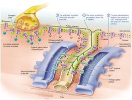
This process links the electrical signal (action potential) to the mechanical event (contraction) in muscle fibers.
Action potential travels down T-tubules.
Triggers Ca2+ release from SR.
Ca2+ enables actin-myosin interaction.
Actin-Myosin Interaction
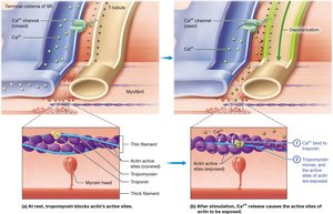
Muscle contraction is regulated by the interaction between actin and myosin, controlled by calcium and regulatory proteins.
At rest: Tropomyosin blocks myosin-binding sites on actin; troponin holds tropomyosin in place.
During contraction: Ca2+ binds troponin, moving tropomyosin and exposing binding sites; myosin binds actin (cross-bridge cycling).
During relaxation: Ca2+ removed; troponin returns tropomyosin to blocking position; cross-bridges detach.
Cross-Bridge Cycling and Role of ATP
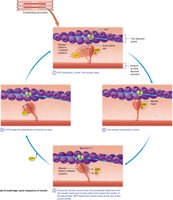
ATP is essential for muscle contraction and relaxation, powering the cross-bridge cycle between actin and myosin.
Myosin binds actin (cross-bridge forms).
Power stroke: Myosin pulls actin, shortening sarcomere.
ATP binds myosin, causing release from actin.
ATP hydrolysis resets myosin to high-energy position.
Myosin ATPase: Enzyme that breaks down ATP, powering contraction and relaxation.
Muscle Twitch and Phases
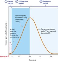
A muscle twitch is a single, brief contraction in response to a stimulus, consisting of three phases.
Latent period: Time between stimulus and contraction.
Contraction phase: Muscle shortens; cross-bridge cycling occurs.
Relaxation phase: Muscle returns to resting length; Ca2+ pumped back into SR.
Tension Production and Summation
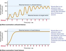
Muscle tension depends on the frequency and pattern of stimulation.
Single twitch: One action potential → one brief contraction.
Summation: Second stimulus before relaxation → increased tension.
Unfused tetanus: Rapid stimuli → partial relaxation → sustained, wavering tension.
Complete tetanus: Very rapid stimuli → no relaxation → maximal, smooth tension.
Motor Units and Muscle Tone
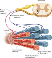
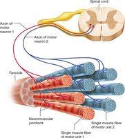
A motor unit consists of a motor neuron and all the muscle fibers it innervates. Muscle tone is the baseline tension maintained by involuntary activation of motor units.
Motor unit: Group of muscle fibers innervated by a single motor neuron.
Muscle tension: Force generated during contraction.
Muscle tone: Small amount of tension at rest.
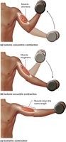
Muscle Contractions: Types
Muscle contractions are classified based on changes in muscle length and tension.
Isotonic: Muscle changes length, tension remains constant; movement occurs.
Concentric: Muscle shortens while generating force.
Eccentric: Muscle lengthens while generating force.
Isometric: Muscle length stays the same, tension changes; no movement.
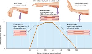
Length-Tension Relationship
The force a muscle can generate depends on the sarcomere length, with optimal overlap of actin and myosin producing maximal tension.
Optimal length: Maximum overlap → maximal tension.
Too short: Excessive overlap → reduced tension.
Too stretched: Minimal overlap → reduced tension.
Motor Unit Recruitment and Muscle Force
Increasing muscle force involves recruiting more motor units and increasing action potential frequency.
Size principle: Smaller motor units activate first; larger units recruited as more force is needed.
Summation: Higher AP frequency increases tension in each motor unit.
Recruitment: More motor units → greater overall force.
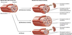
Changes Caused by Physical Training
Physical training induces adaptations in skeletal muscle, affecting its structure and function.
Endurance training: Increases mitochondria, blood vessels, and oxidative enzymes.
Resistance training: Increases myofibril diameter and number, enhancing force.
Disuse: Decreases myofibrils and mitochondria, reducing muscle size and function.
Skeletal Muscle Metabolism
Muscle fibers utilize several metabolic pathways to generate ATP for contraction.
ATP: Immediate energy source for contraction.
Creatine Phosphate (CP): Rapidly regenerates ATP during short, intense activity.
Creatine Kinase (CK): Transfers phosphate from CP to ADP to form ATP.
Glycogen: Stored glucose; substrate for ATP production during sustained activity.
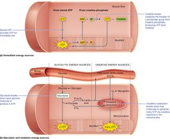
Glycolysis and Oxidative Phosphorylation
Muscle fibers produce ATP via glycolysis (anaerobic) and oxidative phosphorylation (aerobic), depending on activity duration and intensity.
Glycolysis: Occurs in cytosol; anaerobic; produces 2 ATP per glucose quickly; supports short, high-intensity activity.
Oxidative phosphorylation: Occurs in mitochondria; aerobic; produces ~30-32 ATP per glucose slowly; supports long-duration, lower-intensity activity.
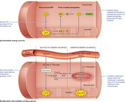
Muscle Fiber Types
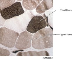
Skeletal muscle fibers are classified based on their metabolic properties, force generation, and fatigue resistance.
Slow Oxidative (Type I): Small diameter, many mitochondria, high myoglobin, fatigue-resistant, aerobic metabolism, low force, used for posture and endurance.
Fast Oxidative (Type IIa): Intermediate diameter, many mitochondria, moderate capillaries and myoglobin, moderately fatigue-resistant, aerobic metabolism, moderate force, used for quick walking and intermediate activities.
Fast Glycolytic (Type IIb): Large diameter, fewer mitochondria, low capillaries and myoglobin, fatigues quickly, anaerobic metabolism, high force, used for short and powerful movements.
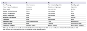
Class | I | IIa | IIb |
|---|---|---|---|
Primary Property | Slow Oxidative | Oxidative Glycolytic | Fast Glycolytic |
Blood Supply | Extensive | Less extensive | Few |
Mitochondria | Large | Intermediate | Small |
Myoglobin | Large | Intermediate | Small |
Fatigability | Low | Intermediate | High |
Diameter | Small | Large | Intermediate |
Example Activities | Standing, sitting | Walking, running | Heavy lifting, sprinting |
Role of Myoglobin in Muscle Fibers
Myoglobin is a protein that binds and stores oxygen inside muscle cells, enhancing aerobic endurance and giving fibers a red color.
Function: Increases oxygen availability for ATP production.
Importance: Contributes to endurance and aerobic metabolism.
