 Back
BackSkeletal Muscle Tissue: Structure, Function, and Types
Study Guide - Smart Notes
 Tailored notes based on your materials, expanded with key definitions, examples, and context.
Skeletal Muscle Tissue
Introduction to Muscle Tissue
Muscle tissue is a fundamental component of the human body, responsible for movement, stability, and heat generation. There are three main types of muscle tissue: skeletal, cardiac, and smooth. Skeletal muscle tissue is the most abundant, making up nearly half of the body’s mass and is primarily involved in voluntary movements.
Properties of Muscle Tissue
Contractility: The ability of muscle cells to shorten, driven by myofilaments composed of actin and myosin.
Excitability: Muscle cells respond to nerve signals, generating electrical impulses along their plasma membrane.
Extensibility: Muscles can be stretched by opposing muscle contractions or by substances within hollow organs.
Elasticity: Muscles recoil to their original length after being stretched.
Muscle Tissue Terminology
Myo and mys: Prefixes meaning "muscle".
Sarco: Prefix meaning "flesh" (e.g., sarcoplasm is the cytoplasm of muscle cells, sarcolemma is the plasma membrane).
Functions of Muscle Tissue
Produce Movement: Skeletal muscles move bones; smooth muscles move substances through organs.
Open and Close Body Passageways: Sphincter muscles act as valves.
Maintain Posture and Stabilize Joints: Muscle tone stabilizes synovial joints.
Heat Generation: Muscle contractions produce heat, helping maintain body temperature.
Types of Muscle Tissue
Skeletal Muscle Tissue
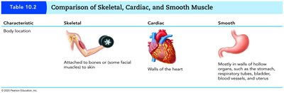
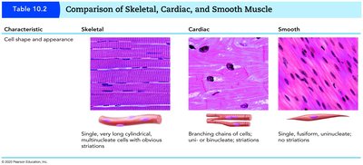
Skeletal muscle tissue is packaged into muscles attached to the skeleton, is striated, and is controlled voluntarily by the nervous system. It constitutes about 40% of body weight.
Cardiac and Smooth Muscle Tissue
Cardiac Muscle: Found only in the heart, striated, involuntary.
Smooth Muscle: Found in walls of hollow organs, non-striated, involuntary.
Gross Anatomy of Skeletal Muscle
Connective Tissue Sheaths and Fascicles
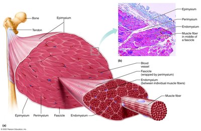
Connective tissue sheaths organize and protect muscle fibers:
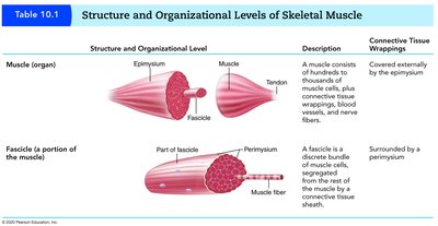
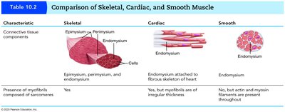
Epimysium: Surrounds the entire muscle.
Perimysium: Surrounds each fascicle (group of muscle fibers).
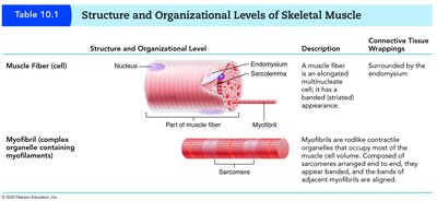
Endomysium: Wraps each individual muscle cell.
Nerves and Blood Vessels
Each skeletal muscle is supplied by one nerve, one artery, and one or more veins. These branches serve individual muscle fibers, ensuring proper function and nourishment.
Muscle Attachments
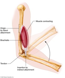
Muscles attach to bones at two points:
Origin: Less movable attachment.
Insertion: More movable attachment.
Attachments can be direct (short connective tissue fibers) or indirect (via tendons or aponeuroses).
Microscopic and Functional Anatomy of Skeletal Muscle Tissue
Skeletal Muscle Fiber Structure
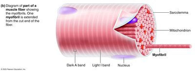
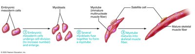
Skeletal muscle fibers are long, cylindrical, multinucleate cells formed by fusion of embryonic cells. Nuclei are located peripherally.
Myofibrils and Sarcomeres
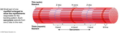
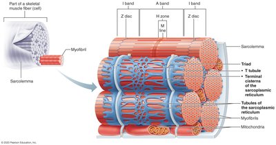
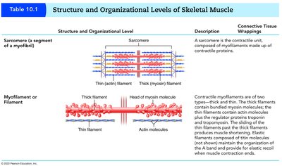
Myofibrils are specialized contractile organelles within muscle fibers, composed of repeating units called sarcomeres. Sarcomeres are the functional units of muscle contraction.
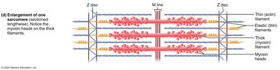
Z disc (Z line): Boundary of each sarcomere.
Thin (actin) filaments: Extend from Z disc toward the center.
Thick (myosin) filaments: Located in the center, overlap with actin filaments.
A band: Full length of thick filament.
H zone: Center of A band with no thin filaments.
I band: Region with only thin filaments.
M line: Center of H zone, holds thick filaments together.
Titin and Other Myofibril Proteins
Titin is a springlike molecule that resists overstretching, holding thick filaments in place and unfolding when muscle is stretched.
Sarcoplasmic Reticulum and T Tubules
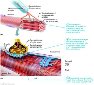
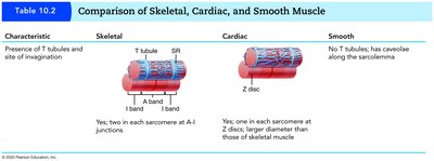
The sarcoplasmic reticulum is a specialized smooth endoplasmic reticulum that surrounds each myofibril and stores calcium ions. T tubules are deep invaginations of the sarcolemma, forming a triad with terminal cisterns of the sarcoplasmic reticulum.
Mechanism of Muscle Contraction
Types of Contraction
Concentric contraction: Muscle shortens to do work.
Eccentric contraction: Muscle generates force as it lengthens, acting as a brake.
Sliding Filament Mechanism
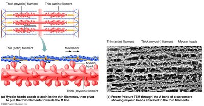
During contraction, myosin heads attach to actin filaments and pull them toward the center of the sarcomere. This process is powered by ATP and initiated by calcium release from the sarcoplasmic reticulum. Thin and thick filaments do not shorten; instead, their overlap increases.
Changes in Striation Pattern During Contraction
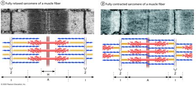
Fully relaxed: Thin filaments partially overlap thick filaments.
Contracted: Z discs move closer, sarcomere shortens, I bands shorten, H zone disappears, A band remains unchanged.
Structure and Organizational Levels of Skeletal Muscle
The structure of skeletal muscle is organized hierarchically from the whole muscle to the molecular level. The following tables summarize these levels:
Innervation of Skeletal Muscle
Neuromuscular Junction
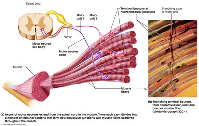
Motor neurons innervate skeletal muscle fibers at the neuromuscular junction. Terminal boutons (axon terminals) store neurotransmitters, which are released into the synaptic cleft to stimulate muscle contraction.
Motor Units
A motor unit consists of a motor neuron and all the muscle fibers it innervates. Each axon branches to form neuromuscular junctions with multiple muscle fibers.
Types of Skeletal Muscle Fibers
Classification by Energy Production and Contraction Speed
Oxidative fibers: Produce ATP aerobically.
Glycolytic fibers: Produce ATP anaerobically via glycolysis.
Three Classes of Skeletal Muscle Fibers
Slow oxidative fibers: Red, rich in myoglobin and mitochondria, contract slowly, resistant to fatigue, small diameter.
Fast glycolytic fibers: White, low myoglobin, few mitochondria, large diameter, contract rapidly, tire quickly, generate more power.
Fast oxidative fibers: Intermediate diameter, contract quickly, oxygen dependent, high myoglobin, somewhat fatigue resistant.
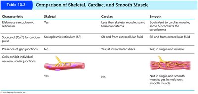
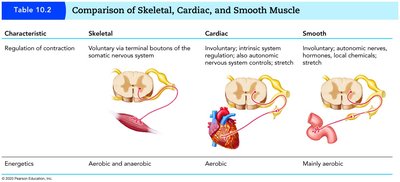
Comparison of Skeletal, Cardiac, and Smooth Muscle
The following tables compare the characteristics of the three muscle types:
Disorders of Muscle Tissue
Muscular Dystrophy
A group of inherited muscle-destroying diseases characterized by muscle enlargement due to fat and connective tissue, followed by degeneration. Types include Duchenne muscular dystrophy and myotonic dystrophy.
Myofascial Pain Syndrome and Fibromyalgia
Myofascial pain syndrome: Caused by tightened bands of muscle fibers.
Fibromyalgia: Chronic pain syndrome with fatigue, sleep abnormalities, and musculoskeletal pain.
Muscle Tissue Throughout Life
Development and Aging
Muscle tissue develops from myoblasts, which fuse to form skeletal muscle fibers. Satellite cells surround fibers and aid in growth. Muscle mass differs between males (42%) and females (36%) due to androgens. With age, connective tissue increases and muscle fibers decrease, leading to sarcopenia (muscle wasting).
Summary Table: Structure and Organizational Levels of Skeletal Muscle
Level | Description | Connective Tissue Wrapping |
|---|---|---|
Muscle (organ) | Thousands of muscle cells, plus connective tissue, blood vessels, and nerves | Epimysium |
Fascicle | Discrete bundle of muscle cells | Perimysium |
Muscle Fiber (cell) | Elongated, multinucleate cell with striated appearance | Endomysium |
Myofibril | Rodlike contractile organelle, composed of sarcomeres | None |
Sarcomere | Contractile unit, composed of myofilaments | None |
Myofilament | Actin (thin) and myosin (thick) filaments | None |
Key Equations
ATP Hydrolysis:
Force Generation:
