 Back
Skeletal Muscles: Origins, Insertions, and Actions
Study Guide - Smart Notes
 Tailored notes based on your materials, expanded with key definitions, examples, and context.
Skeletal Muscles: Overview
Introduction to Skeletal Muscles
Skeletal muscles are voluntary muscles attached to bones and are responsible for movement, posture, and stability. Each muscle has a specific origin (attachment point), insertion (end point), and action (movement produced). Understanding these features is essential for identifying muscles and their functions.
Origin: The fixed attachment point of the muscle, usually proximal.
Insertion: The movable attachment point, usually distal.
Action: The movement produced when the muscle contracts.
Axial Skeletal Muscles
Anterior Axial Muscles
The anterior muscles of the axial skeleton are primarily involved in facial expression, head movement, and respiration.
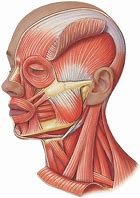
Orbicularis Oculi: Origin: Frontal and Maxilla bones Insertion: Skin of the eyelids Action: Closes the eyelids
Orbicularis Oris: Origin: Maxilla and Mandible Insertion: Skin of the lips Action: Closes/Purses the lips ("kissing muscle")
Masseter: Origin: Zygomatic arch Insertion: Mandibular ramus Action: Elevates jaw
Sternocleidomastoid: Origin: Manubrium, clavicle Insertion: Mastoid process Action: Flex and laterally rotate head
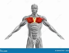
Pectoralis Major: Origin: Clavicle, sternum Insertion: Greater tubercle of humerus Action: Flex, adduct, and medially rotate arm
Serratus Anterior: Origin: Ribs 1-8 Insertion: Medial border of scapula Action: Rotates scapula
Intercostal Muscles
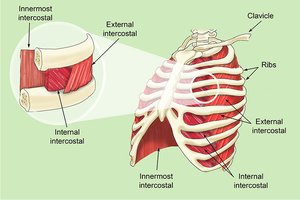
Intercostal muscles are located between the ribs and play a crucial role in breathing.
Internal Intercostals: Origin: Superior border of inferior rib Insertion: Inferior border of superior rib Action: Depresses ribcage during exhalation
External Intercostals: Origin: Inferior border of superior rib Insertion: Superior border of inferior rib Action: Elevates ribcage during inhalation
Abdominal Muscles
The abdominal muscles support trunk movement and stability.
Rectus Abdominis: Origin: Pubic symphysis Insertion: Xiphoid process, costal cartilage Action: Flex and rotate lumbar spine
External Oblique: Origin: Lower 8 ribs Insertion: Linea alba Action: Trunk rotation
Posterior Axial Muscles
The posterior muscles of the axial skeleton are involved in movements of the back, shoulders, and hips.
Trapezius: Origin: Occipital bone, spinous processes of C7-T12 Insertion: Acromion process and scapular spine Action: Raise, retract, and rotate scapular spine
Latissimus Dorsi: Origin: Thoracic and lumbar vertebrae, ribs, iliac crest Insertion: Proximal humerus Action: Extend, adduct, and medially rotate arm
Gluteus Maximus: Origin: Sacrum, coccyx, ilium Insertion: Iliotibial tract Action: Extends thigh
Summary Table: Muscle Origins, Insertions, and Actions
Muscle | Origin | Insertion | Action |
|---|---|---|---|
Orbicularis Oculi | Frontal & Maxilla bones | Skin of eyelids | Closes eyelids |
Orbicularis Oris | Maxilla & Mandible | Skin of lips | Closes/Purses lips |
Masseter | Zygomatic arch | Mandibular ramus | Elevates jaw |
Sternocleidomastoid | Manubrium, clavicle | Mastoid process | Flex & rotate head |
Pectoralis Major | Clavicle, sternum | Greater tubercle of humerus | Flex, adduct, medially rotate arm |
Serratus Anterior | Ribs 1-8 | Medial border of scapula | Rotates scapula |
Internal Intercostals | Superior border of inferior rib | Inferior border of superior rib | Depresses ribcage |
External Intercostals | Inferior border of superior rib | Superior border of inferior rib | Elevates ribcage |
Rectus Abdominis | Pubic symphysis | Xiphoid process, costal cartilage | Flex & rotate lumbar spine |
External Oblique | Lower 8 ribs | Linea alba | Trunk rotation |
Trapezius | Occipital bone, C7-T12 | Acromion process, scapular spine | Raise, retract, rotate scapula |
Latissimus Dorsi | Thoracic/lumbar vertebrae, ribs, iliac crest | Proximal humerus | Extend, adduct, medially rotate arm |
Gluteus Maximus | Sacrum, coccyx, ilium | Iliotibial tract | Extends thigh |
Deltoid | Clavicle, acromion process | Diaphysis of humerus | Arm abduction |
Biceps Brachii | Coracoid process | Radial tuberosity | Elbow flexion |
Brachialis | Distal humerus | Coronoid process | Lifts ulna with elbow flexion |
Brachioradialis | Distal humerus | Styloid process of radius | Lifts radius with elbow flexion |
Tensor Fascia Latae | Iliac crest | Iliotibial tract | Steadies knee |
Rectus Femoris | Anterior iliac spine | Patella, tibial tuberosity | Extend knee, flex thigh |
Vastus Lateralis | Greater trochanter | Patella, tibial tuberosity | Extend knee, flex thigh |
Vastus Medialis | Diaphysis of femur | Patella, tibial tuberosity | Extend knee, flex thigh |
Sartorius | Anterior iliac spine | Proximal tibia | Flex, abduct, laterally rotate thigh |
Gracilis | Pubis, ischium | Medial tibia | Adduct thigh |
Tibialis Anterior | Tibia | Medial cuneiform, 1st metatarsal | Dorsiflexion |
Triceps Brachii | Posterior humerus | Olecranon process | Extends elbow |
Biceps Femoris | Ischial tuberosity | Fibula | Flex knee, extend thigh |
Semitendinosus | Ischial tuberosity | Tibia | Flex knee, extend thigh |
Semimembranosus | Ischial tuberosity | Tibia | Flex knee, extend thigh |
Gastrocnemius | Distal femur | Calcaneus | Plantar flexion |
Soleus | Fibula, tibia | Calcaneus | Plantar flexion |
Key Concepts and Applications
Muscle identification: Recognizing muscles by their origin, insertion, and action is essential for anatomy and physiology studies.
Clinical relevance: Understanding muscle actions helps diagnose injuries and plan rehabilitation.
Movement analysis: Muscles work in groups to produce complex movements; knowing their functions aids in understanding biomechanics.
Example: The sternocleidomastoid muscle is often palpated during physical exams to assess neck movement and flexibility. Additional info: Muscles are often named based on their location, shape, or function (e.g., deltoid for its triangular shape, pectoralis major for its location in the chest).
