 Back
BackSkeletal Overview and the Axial Skeleton: Study Notes
Study Guide - Smart Notes
 Tailored notes based on your materials, expanded with key definitions, examples, and context.
Skeletal Overview
Classification and Structure of Bone & Cartilages
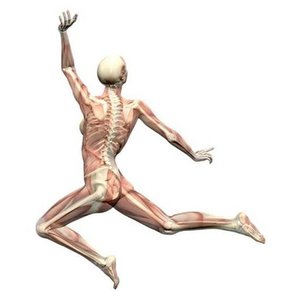
The human skeleton is constructed from two of the most supportive tissues: bone and cartilage. Bone forms the framework of the body, providing support and protection for organ systems, while cartilage is found in areas requiring flexibility and resilience.
Bone Functions:
Provides a lever system for movement in conjunction with skeletal muscles
Acts as a storage depot for lipids and minerals (e.g., calcium, phosphate)
Site for hematopoiesis (blood cell formation) in the marrow
Cartilage Functions:
Provides flexibility and resilience in specific locations (e.g., joints, rib cage)
Joints (Articulations): Bones are connected at joints, allowing movement and stability.
Classification of Bone
Bones are classified by their shape and structure.
Shape (Gross Anatomy):
Long bones (e.g., femur, humerus)
Short bones (e.g., carpals, tarsals)
Flat bones (e.g., skull, sternum)
Irregular bones (e.g., vertebrae, pelvis)
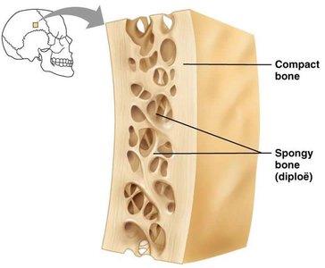
Structure (Texture):
Compact bone: Dense, strong outer layer
Spongy (cancellous) bone: Porous, inner layer, often found at the ends of long bones and inside flat bones
The Skeleton
Divisions of the Skeleton
The skeleton is subdivided into two main divisions:
Axial Skeleton: Composed of the skull, vertebral column, and thoracic cage
Appendicular Skeleton: Includes the limbs and girdles (not covered in detail here)
Bone Markings
Types and Functions of Bone Markings
Bones are not smooth; they have various projections, depressions, ridges, and holes called bone markings. These serve as sites for muscle attachment, conduits for nerves and blood vessels, and help form joints.
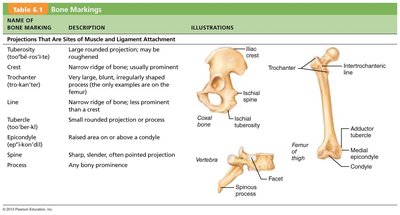
Projections: Sites for muscle and ligament attachment (e.g., tuberosity, crest, trochanter)
Depressions and Openings: Allow passage of blood vessels and nerves (e.g., foramen, fossa)
Joint Formation: Markings that help form joints (e.g., condyle, head)
Name of Bone Marking | Description | Illustrations |
|---|---|---|
Tuberosity | Large rounded projection; may be roughened | Illustrated on ilium and femur |
Crest | Narrow ridge of bone; usually prominent | Illustrated on ilium |
Trochanter | Very large, blunt, irregularly shaped process | Illustrated on femur |
Line | Narrow ridge of bone; less prominent than a crest | Illustrated on femur |
Tubercle | Small rounded projection or process | Illustrated on femur |
Epicondyle | Raised area on or above a condyle | Illustrated on femur |
Spine | Sharp, slender, often pointed projection | Illustrated on vertebra |
Process | Any bony prominence | Illustrated on vertebra |
The Axial Skeleton
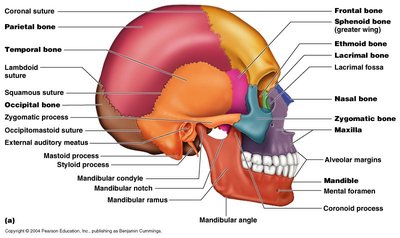
Skull
The skull is a complex structure composed of several bones joined by sutures. It protects the brain and forms the structure of the face.
Major Bones: Frontal, parietal, temporal, occipital, sphenoid, ethmoid, nasal, zygomatic, maxilla, mandible
Sutures: Immovable joints between skull bones (e.g., coronal, sagittal, lambdoid, squamous)
Bone Markings: Processes, condyles, and foramina for muscle attachment and passage of nerves/vessels
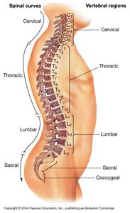
Vertebral Column
The vertebral column (spine) is composed of individual vertebrae and provides support, protection for the spinal cord, and flexibility.
Regions:
Cervical (7 vertebrae, C1–C7)
Thoracic (12 vertebrae, T1–T12)
Lumbar (5 vertebrae, L1–L5)
Sacral (5 fused vertebrae, sacrum)
Coccygeal (3–5 fused vertebrae, coccyx)
Spinal Curves: Cervical and lumbar curves are concave; thoracic and sacral curves are convex
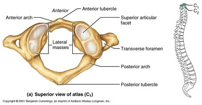
Special Cervical Vertebrae: Atlas and Axis
The first two cervical vertebrae are highly modified to allow special functions.
Atlas (C1): Supports the skull, allows nodding motion
Axis (C2): Has the odontoid process (dens), allows rotation of the head
Unique Features: Lateral masses, anterior and posterior arches, transverse foramen
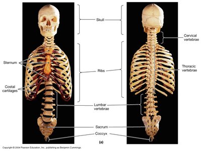
Thoracic Cage
The thoracic cage consists of the ribs, sternum, and thoracic vertebrae. It protects vital organs (heart, lungs) and supports the shoulder girdles and upper limbs.
Rib Articulation: Ribs articulate with thoracic vertebrae and the sternum via costal cartilages
Components: Sternum, ribs (true, false, floating), costal cartilages
Example: The rib cage expands during inhalation to allow lung expansion.
