 Back
BackSkeletal System and Axial Skeleton: Structure, Classification, and Anatomy
Study Guide - Smart Notes
 Tailored notes based on your materials, expanded with key definitions, examples, and context.
Skeletal System Overview
Structure of Bones
The skeletal system is composed of various types of bones, each with unique structural features that contribute to their function. Bones are classified based on their shape and internal structure, which is essential for understanding their roles in the body.
Long Bones: Characterized by a shaft (diaphysis), two ends (epiphyses), and a medullary cavity. Example: femur.
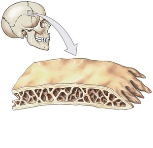
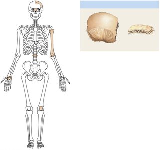
Flat Bones: Consist of a layer of spongy bone (diploë) sandwiched between two layers of compact bone. Example: parietal bone of the skull.
Example: The femur is a typical long bone, while the parietal bone is a flat bone.
Bone Histology
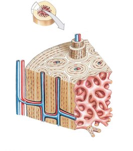
Bones are composed of compact and spongy tissue, each with distinct histological features. Compact bone is organized into osteons, while spongy bone consists of trabeculae.
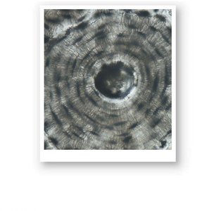
Osteon: The structural unit of compact bone, consisting of concentric lamellae surrounding a central canal.
Lamellae: Layers of bone matrix within osteons.
Canaliculi: Small channels that connect lacunae and allow for nutrient and waste exchange.
Trabeculae: The lattice-like network in spongy bone, providing structural support and space for marrow.
Example: Compact bone is found in the diaphysis of long bones, while spongy bone is prominent in the epiphyses and flat bones.
Classification of Bones
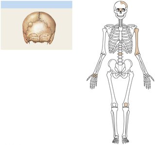
Types of Bones by Shape
Bones are classified into several types based on their shape and location in the skeleton. This classification aids in understanding their function and anatomical relationships.
Sutural Bones: Small, irregular bones found within sutures of the skull.
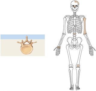
Irregular Bones: Complex shapes, such as vertebrae.
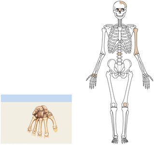
Short Bones: Cube-shaped bones, such as carpals.
Flat Bones: Thin, parallel surfaces, such as the parietal bone.
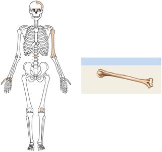
Long Bones: Longer than they are wide, such as the humerus.
Sesamoid Bones: Small, round bones embedded in tendons, such as the patella.
Example: The vertebra is an irregular bone, the carpal bones are short bones, and the patella is a sesamoid bone.
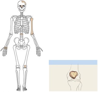
Bone Markings and Features
Bone Markings
Bones display various markings that serve as sites for muscle attachment, passageways for nerves and blood vessels, and articulations with other bones. Understanding these markings is crucial for anatomical identification and clinical relevance.
Openings: Foramen, canal, fissure, meatus, sinus.
Elevations and Projections: Process, ramus, head, trochanter, crest, spine, tubercle, tuberosity, facet, condyle, trochlea.
Depressions: Sulcus, fossa.
Example: The foramen magnum in the occipital bone allows passage of the spinal cord.
Axial Skeleton
Components of the Axial Skeleton
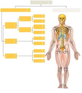
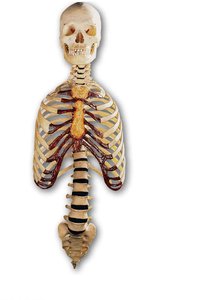
The axial skeleton forms the central axis of the body and consists of the skull, vertebral column, and thoracic cage. It provides support and protection for the brain, spinal cord, and thoracic organs.
Skull: Composed of cranial and facial bones.
Vertebral Column: Includes cervical, thoracic, lumbar, sacral, and coccygeal regions.
Thoracic Cage: Consists of the sternum and ribs.
Example: The axial skeleton contains 80 bones, including the cranium, vertebrae, and ribs.
Skull Anatomy
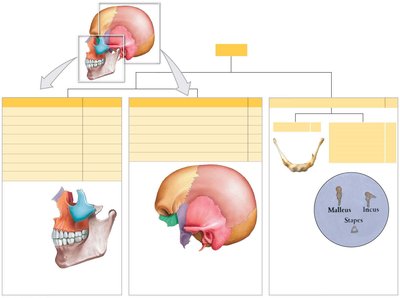
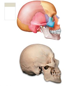
The skull is divided into cranial and facial subdivisions, each with specific bones and functions. Cranial bones protect the brain, while facial bones support the entrances to the digestive and respiratory tracts.
Cranial Bones: Frontal, parietal, temporal, occipital, sphenoid, ethmoid.
Facial Bones: Maxillae, palatine, nasal, inferior nasal conchae, zygomatic, lacrimal, vomer, mandible.
Example: The mandible is the only movable bone of the skull.
Major Sutures of the Skull
Sutures are immovable joints between skull bones. Major sutures include the coronal, sagittal, lambdoid, and squamous sutures.
Coronal Suture: Between frontal and parietal bones.
Sagittal Suture: Between parietal bones.
Lambdoid Suture: Between parietal and occipital bones.
Squamous Suture: Between parietal and temporal bones.
Example: The lambdoid suture is located at the posterior aspect of the skull.
Vertebral Column
Regions and Curves of the Vertebral Column
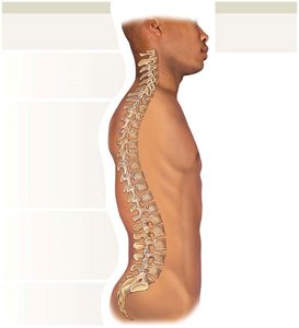
The vertebral column is divided into five regions, each with characteristic vertebrae. It exhibits four curves: cervical, thoracic, lumbar, and sacral, which help balance and support the body.
Cervical (7 vertebrae): Supports the head.
Thoracic (12 vertebrae): Supports the thoracic cage.
Lumbar (5 vertebrae): Supports the lower back.
Sacral (5 fused vertebrae): Forms the sacrum.
Coccygeal (3-5 fused vertebrae): Forms the coccyx.
Example: The cervical curve develops as infants learn to balance their head.
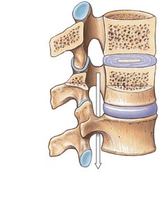
Vertebral Anatomy
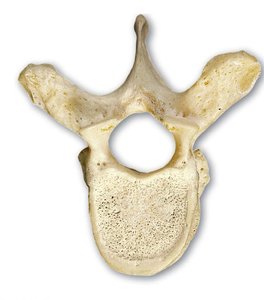
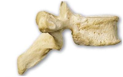
Each vertebra has a vertebral body, arch, and several processes. The anatomy varies by region, reflecting functional differences.
Vertebral Body: Main weight-bearing portion.
Vertebral Arch: Encloses the vertebral foramen.
Processes: Spinous, transverse, and articular processes for muscle attachment and articulation.
Example: Thoracic vertebrae have facets for rib articulation.
Thoracic Cage
Structure and Function of the Thoracic Cage
The thoracic cage protects vital organs and supports the upper body. It consists of the sternum and ribs, which are classified as true, false, and floating ribs.
Sternum: Manubrium, body, xiphoid process.
Ribs: 12 pairs; ribs 1-7 are true ribs, 8-10 are false ribs, 11-12 are floating ribs.
Example: True ribs attach directly to the sternum via costal cartilage.
Summary Table: Bone Classification
Bone Type | Example | Location |
|---|---|---|
Long Bone | Femur | Limbs |
Short Bone | Carpals | Wrist |
Flat Bone | Parietal | Skull |
Irregular Bone | Vertebra | Spine |
Sesamoid Bone | Patella | Knee |
Sutural Bone | Small bones in skull sutures | Skull |
