 Back
BackSkeletal System: Bone Tissue, Bones, and Joints – Study Notes
Study Guide - Smart Notes
 Tailored notes based on your materials, expanded with key definitions, examples, and context.
Skeletal System: Bone Tissue, Bones, and Joints
Introduction to the Skeletal System
The skeletal system provides the structural framework for the human body, protects vital organs, facilitates movement, stores minerals, and houses bone marrow for blood cell production. This unit covers the structure and function of bone tissue, the anatomy of bones, and the classification and function of joints.
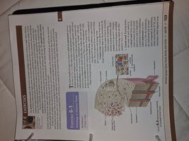
Bone Tissue
Structure and Function of Bone Tissue
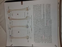
Bone tissue is a specialized connective tissue composed of cells, fibers, and ground substance.
It provides support, protection, and enables movement by serving as points of attachment for muscles.
Bone tissue is dynamic, constantly being remodeled through the actions of osteoblasts (bone-forming cells) and osteoclasts (bone-resorbing cells).
There are two main types of bone tissue: compact bone (dense and strong) and spongy bone (porous and lightweight).
Axial Skeleton
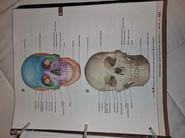
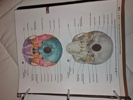
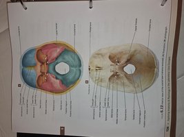
Skull Anatomy
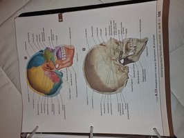
The skull is a complex structure composed of cranial and facial bones. It protects the brain and forms the structure of the face.
Cranial bones include the frontal, parietal, temporal, occipital, sphenoid, and ethmoid bones.
Facial bones include the maxilla, mandible, zygomatic, nasal, lacrimal, palatine, inferior nasal concha, and vomer.
Sutures are immovable joints that connect the bones of the skull.

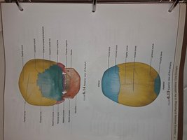
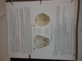
Infant Skull and Fontanelles
Infant skulls have soft spots called fontanelles that allow for growth of the brain and skull during development.
Fontanelles ossify and become sutures as the child grows.
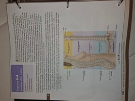
Vertebral Column
The vertebral column, or spine, supports the head and trunk, protects the spinal cord, and provides attachment points for ribs and muscles.
It consists of 33 vertebrae in five regions: cervical (7), thoracic (12), lumbar (5), sacral (5 fused), and coccygeal (4 fused).
Intervertebral discs act as shock absorbers between vertebrae.
Appendicular Skeleton
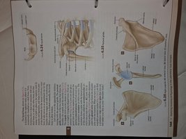
Pectoral Girdle and Upper Limb
The pectoral girdle consists of the clavicle and scapula, connecting the upper limb to the axial skeleton.
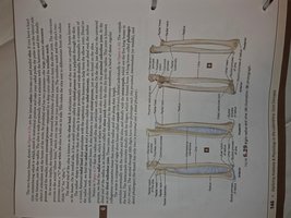
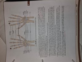
The upper limb includes the humerus, radius, ulna, carpals, metacarpals, and phalanges.
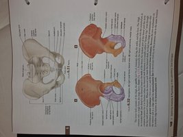
Pelvic Girdle and Lower Limb
The pelvic girdle consists of two hip bones (coxal bones), which articulate with the sacrum and femur.
The lower limb includes the femur, patella, tibia, fibula, tarsals, metatarsals, and phalanges.
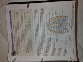
Thoracic Cage
The thoracic cage is formed by the sternum, ribs, and thoracic vertebrae.
It protects the heart and lungs and supports the shoulder girdles and upper limbs.
There are 12 pairs of ribs: true ribs (1–7), false ribs (8–12), and floating ribs (11–12).
Joints (Articulations)
Classification of Joints
Fibrous joints: Immovable joints held together by fibrous tissue (e.g., sutures of the skull).
Cartilaginous joints: Slightly movable joints connected by cartilage (e.g., intervertebral discs).
Synovial joints: Freely movable joints with a synovial cavity (e.g., shoulder, knee, elbow).
Structure of Synovial Joints
Synovial joints have articular cartilage, a joint (synovial) cavity, an articular capsule, synovial fluid, reinforcing ligaments, and nerves and blood vessels.
Types of synovial joints include hinge, pivot, ball-and-socket, saddle, condyloid, and plane joints.
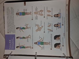
Movements at Synovial Joints
Movements include flexion, extension, abduction, adduction, rotation, circumduction, pronation, and supination.

Summary Table: Major Bones of the Human Skeleton
Region | Major Bones |
|---|---|
Skull | Frontal, Parietal, Temporal, Occipital, Sphenoid, Ethmoid, Maxilla, Mandible, Zygomatic |
Vertebral Column | Cervical, Thoracic, Lumbar, Sacrum, Coccyx |
Thoracic Cage | Sternum, Ribs |
Pectoral Girdle | Clavicle, Scapula |
Upper Limb | Humerus, Radius, Ulna, Carpals, Metacarpals, Phalanges |
Pelvic Girdle | Hip bones (Ilium, Ischium, Pubis) |
Lower Limb | Femur, Patella, Tibia, Fibula, Tarsals, Metatarsals, Phalanges |
Additional info:
Bone is a reservoir for minerals, especially calcium and phosphate.
Red bone marrow is the site of hematopoiesis (blood cell formation).
Osteoporosis is a common disorder characterized by decreased bone mass and increased fracture risk.
