 Back
BackSkeletal System: Structure, Function, and Joints
Study Guide - Smart Notes
 Tailored notes based on your materials, expanded with key definitions, examples, and context.
Skeletal System Overview
Main Organs and Functions
The skeletal system is a supportive connective tissue system that provides the framework for the body, protects internal organs, stores minerals and fat, produces blood cells, and enables movement through articulation with muscles. It consists of bones, cartilages, and ligaments.
Bones: Provide structure, protection, and serve as levers for movement.
Cartilage: Offers flexible support, reduces friction at joints, and forms the template for bone development.
Ligaments: Connect bone to bone, stabilizing joints.
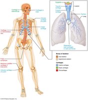
Cartilage: Structure and Function
Types and Roles of Cartilage
Cartilage is a resilient and smooth elastic tissue, a rubber-like padding that covers and protects the ends of long bones at the joints and is a structural component of the rib cage, ear, nose, bronchial tubes, and intervertebral discs.
Functions: Provides support, flexibility, and smooth surfaces for joint movement; forms the embryonic skeleton; and assists in bone growth.
Types: Hyaline cartilage, elastic cartilage, and fibrocartilage.
Matrix: Composed of glycosaminoglycans (GAGs), proteoglycans, collagen, and water.
Cells: Chondroblasts (produce matrix) and chondrocytes (maintain matrix).
Comparison of Cartilage and Bone
Structural and Functional Differences
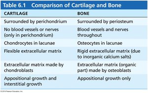
Cartilage and bone are both connective tissues but differ in structure, composition, and function.
Cartilage | Bone |
|---|---|
Surrounded by perichondrium | Surrounded by periosteum |
No blood vessels or nerves (except in perichondrium) | Blood vessels and nerves throughout |
Chondrocytes in lacunae | Osteocytes in lacunae |
Flexible extracellular matrix | Rigid extracellular matrix (due to inorganic calcium salts) |
Matrix made by chondroblasts | Matrix (organic part) made by osteoblasts |
Appositional and interstitial growth | Appositional growth only |
Classification of Bones
By Shape and Structure
Bones are classified based on their shape and location in the body.
By Shape:
Long bones (e.g., femur, humerus)
Short bones (e.g., carpals, tarsals)
Flat bones (e.g., sternum, skull)
Irregular bones (e.g., vertebrae, pelvis)
By Structure: Compact (dense) and cancellous (spongy) bone.
Bone Cells
Types and Functions
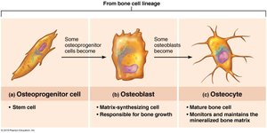
Bone tissue contains four main types of specialized cells, each with distinct roles in bone formation, maintenance, and remodeling.
Osteogenic cells: Stem cells from periosteum and endosteum; differentiate into osteoblasts.
Osteoblasts: Bone-forming cells; secrete bone matrix (osteoid).
Osteocytes: Mature bone cells; maintain bone matrix and monitor mechanical stress.
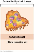
Osteoclasts: Bone-resorbing cells; break down bone matrix, derived from white blood cell lineage.
Bone Matrix
Composition and Properties
The bone matrix is composed of inorganic and organic components, providing both hardness and flexibility.
Inorganic: Hydroxyapatite (calcium phosphate crystals) – gives hardness and strength.
Organic: Collagen fibers, chondroitin sulfate, hyaluronic acid, and water – provide toughness and flexibility.
Anatomy of a Long Bone
Regions and Layers
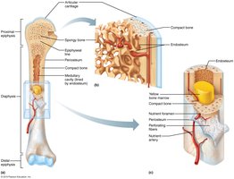
Long bones have distinct regions and layers, each with specific functions in growth, support, and nutrient supply.
Regions: Epiphysis (ends), diaphysis (shaft), epiphyseal line (growth plate remnant).
Layers (superficial to deep): Periosteum (fibrous and osteogenic layers), compact bone, spongy bone, medullary cavity (contains marrow).
Microscopic Structure of Bone
Compact vs. Spongy Bone
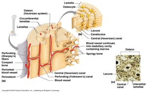
Bone tissue is organized into compact and spongy types, each with unique microscopic features.
Compact bone: Contains osteons (Haversian systems) with concentric lamellae, central canals, and canaliculi for nutrient delivery.
Spongy bone: Composed of trabeculae; lacks central canals; nutrients diffuse through canaliculi.
Bone Formation (Ossification)
Intramembranous and Endochondral Ossification
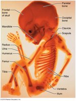
Bone develops through two main processes: intramembranous and endochondral ossification.
Intramembranous ossification: Osteoblasts form bone directly from mesenchymal tissue (e.g., skull, clavicle).
Endochondral ossification: Bone forms by replacing hyaline cartilage (e.g., long bones, axial skeleton).
Epiphyseal Plate and Bone Growth
Zones and Hormonal Regulation
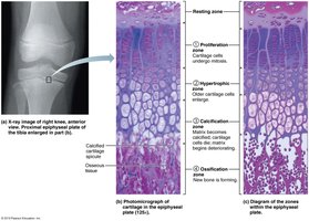
The epiphyseal plate enables longitudinal bone growth through distinct cellular zones. Growth is regulated by hormones such as growth hormone, sex hormones, thyroxine, and insulin.
Zones: Reserve cartilage, proliferation, hypertrophy, calcification, ossification.
Hormones: Stimulate or inhibit growth and closure of the growth plate.
Bone Remodeling and Calcium Regulation
Dynamic Process and Hormonal Control
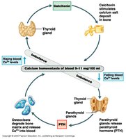
Bone remodeling is a lifelong process involving osteoblast and osteoclast activity. Calcium homeostasis is tightly regulated by hormones.
Parathyroid hormone (PTH): Increases blood calcium by stimulating osteoclasts and increasing intestinal absorption.
Calcitonin: Lowers blood calcium by inhibiting osteoclasts and promoting calcium deposition in bone.
Calcitriol (Vitamin D): Enhances calcium absorption from the gut.
Fractures and Bone Healing
Types and Stages of Healing
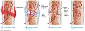
Fractures are breaks in bone continuity, classified as simple, compound, comminuted, or greenstick. Healing occurs in four stages:
Hematoma formation (2 days)
Soft callus formation (2 weeks)
Hard callus formation (2 months)
Remodeling (up to 2 years)
Joints (Articulations)
Definition and Classification
Joints are sites where two or more bones meet. They are classified by structure and function.
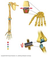
Functional classification: Synarthroses (immovable), amphiarthroses (slightly movable), diarthroses (freely movable).
Structural classification: Fibrous, cartilaginous, and synovial joints.
Fibrous Joints
Types and Examples
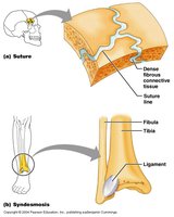
Fibrous joints are connected by dense connective tissue and allow little or no movement.
Sutures: Found in the skull; immovable.
Syndesmoses: Bones joined by ligaments; slight movement (e.g., distal tibiofibular joint).
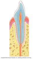
Gomphoses: Peg-in-socket joints (e.g., teeth in jaw).
Cartilaginous Joints
Types and Examples
Cartilaginous joints are united by cartilage and allow limited movement.
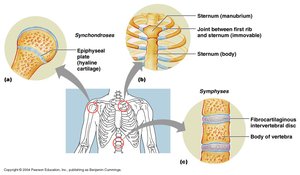
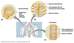
Symphyses: Bones joined by fibrocartilage (e.g., intervertebral discs, pubic symphysis).
Synchondroses: Bones joined by hyaline cartilage (e.g., epiphyseal plate, first rib-sternum joint).
Synovial Joints
Characteristics and Types
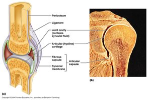
Synovial joints are the most movable type of joint, characterized by a joint cavity filled with synovial fluid, articular cartilage, a fibrous capsule, and supporting ligaments.
Types: Plane (gliding), hinge, pivot, condylar (ellipsoid), saddle, ball-and-socket.
Features: Menisci, bursae, fat pads for shock absorption and stability.
Movements at Synovial Joints
Types of Movement
Synovial joints allow a variety of movements, including gliding, angular, and rotational movements.
Movement | Definition |
|---|---|
Gliding | Sliding flat surfaces of two bones across each other |
Flexion | Decreasing the angle between two bones |
Extension | Increasing the angle between two bones |
Abduction | Moving a limb away from the body midline |
Adduction | Moving a limb toward the body midline |
Circumduction | Moving a limb or finger so that it describes a cone in space |
Rotation | Turning a bone around its longitudinal axis |
Medial rotation | Rotating toward the median plane |
Lateral rotation | Rotating away from the median plane |
Major Synovial Joints
Examples and Features
Temporomandibular Joint (TMJ): Combination plane/ellipsoid; allows elevation, depression, protraction, retraction; contains fibrocartilage disc.
Shoulder (Glenohumeral) Joint: Ball-and-socket; stabilized by rotator cuff muscles and ligaments; allows wide range of motion.
Elbow Joint: Hinge joint; stabilized by collateral and annular ligaments.
Hip (Coxal) Joint: Ball-and-socket; very stable due to deep socket and strong ligaments.
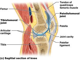
Knee Joint: Largest, most complex; hinge with some rotation; stabilized by menisci, cruciate and collateral ligaments, and bursae.
Joint Disorders
Common Pathologies
Dislocations: Bones forced out of alignment (e.g., shoulder, fingers).
Osteoarthritis: Degeneration of articular cartilage due to wear and tear; non-inflammatory.
Rheumatoid arthritis: Autoimmune inflammation of synovial membrane, leading to cartilage destruction.
